Download

1 / 56
560 likes | 572 Views
Learn about the different stages, clinical types, diagnosis, and treatment of Leishmaniasis, as well as the African sleeping sickness caused by Trypanosoma parasites. Explore the life cycles, geographical distribution, and complications associated with these diseases.

E N D
Promastigotes of Leishmania Amastigote of Leishmania
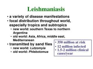
Clinical types of cutaneous leishmaniasis • Leishmania major:Zoonotic cutaneous leishmaniasis: wet lesions with severe reaction • Leishmania tropica:Anthroponotic cutaneous leishmaniasis: Dry lesions with minimal ulceration Oriental sore (most common) classical self-limited ulcer
Uncommon types • Diffuse cutaneous leishmaniasis (DCL): Caused by L. aethiopica, diffuse nodular non-ulcerating lesions. Low immunity to Leishmania antigens, numerous parasites. • Leishmaniasis recidiva (lupoid leishmaniasis): Severe immunological reaction to leishmania antigen leading to persistent dry skin lesions, few parasites.
Diffuse cutaneous leishmaniasis Leishmaniasis recidiva
cutaneous leishmaniasis Diagnosis: • Smear: Giemsa stain – microscopy for LD bodies (amastigotes) • Biopsy: microscopy for LD bodies or culture in NNN medium for promastigotes
Treatment • No treatment – self-healing lesions • Medical: • Pentavalent antimony (Pentostam), Amphotericin B • +/- Antibiotics for secondary bacterial infection. • Surgical: • Cryosurgery • Excision • Curettage
Pentostam ( sodium stibogluconate) for treatment of all types of leishmaniasis
Visceral leishmaniasis • There are geographical variations. • The diseases is called kala-azar • Leishmania infantum mainly affect children • Leishmania donovani mainly affects adults
Presentation • Fever • Splenomegaly, hepatomegaly, hepatosplenomegaly • Weight loss • Anaemia • Epistaxis • Cough • Diarrhoea
Untreated disease can be fatal After recovery it might produce a condition called post kala-azar dermal leishmaniasis (PKDL)
Visceral leishmaniasis Diagnosis • Parasitological diagnosis: METHOD Bone marrow aspirate 1. microscopy Splenic aspirate 2. culture in NNNmedium Lymph node Tissue biopsy
Bone marrow aspiration Bone marrow amastigotes
(2) Immunological Diagnosis: • Specific serologic tests: Direct Agglutination Test (DAT), ELISA, IFAT • Skin test (leishmanin test) for survey of populations and follow-up after treatment. • Non specific detection of hypergammaglobulinaem by formaldehyde (formol-gel) test or by electrophoresis.
DAT test ELISA test
Treatment: • Pentavalent antimony- sodium stibogluconate (Pentostam) • Amphotericin B Treatment of complications: • Anaemia • Bleeding • Infections etc.
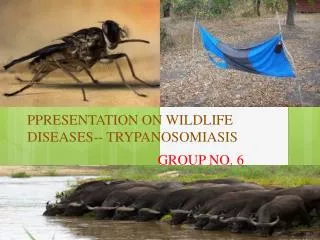
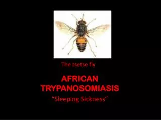
African Trypanosomiasis Life cycle of Trypanosoma brucei gambiense & T. b. rhodesiense
African sleeping sickness Trypanosoma brucei rhodesiense: East Africa, wild and domestic animal reservoirs Trypanosoma brucei gambiense: West and Central Africa, mainly human infection
Pathology and clinical picture • Skin stage: chancre. • Haematolymphatic stage: generalized lymphadenopathy, anaemia, generalized organ involvement. • Central nervous system stage (CNS): Meningoencephalitis. (Development of the disease more rapid in Trypanosoma brucei rhodesiense)
AMERICAN TRYPANOSOMIASIS LIFE CYCLE OF Trypanosoma cruzi