Download
1 / 182
1.84k likes | 2.07k Views
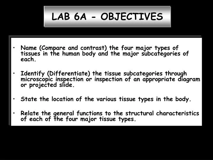
LAB 6A - OBJECTIVES. Name (Compare and contrast) the four major types of tissues in the human body and the major subcategories of each. Identify (Differentiate) the tissue subcategories through microscopic inspection or inspection of an appropriate diagram or projected slide.

E N D
LAB 6A - OBJECTIVES • Name (Compare and contrast) the four major types of tissues in the human body and the major subcategories of each. • Identify (Differentiate) the tissue subcategories through microscopic inspection or inspection of an appropriate diagram or projected slide. • State the location of the various tissue types in the body. • Relate the general functions to the structural characteristics of each of the four major tissue types.
Name the four major types of tissues in the human body and the major subcategories of each. • Epithelial • Connective • Muscle 4. Nervous All have distinctive structures patterns functions
Name the four major types of tissues in the human body and the major subcategories of each. • Epithelial • Classification based on • cell shape • Squamous - width of the cell • is greater than its height • Cuboidal - width, depth, and • height are approximately equal • Columnar - height appreciably • exceeds its width
Name the four major types of tissues in the human body and the major subcategories of each. • Epithelial Classification based on number of layers In a stratified epithelium, the shape of the cells forming the surface layer is used in classifying the epithelium.
Epithelial Tissue • Cellularity – composed almost entirely of close-packed cells • Specialized contacts – tight junctions and desmosomes • Polarity – apical (free surface to which no cellular or extracellular elements adhere) and basal surfaces • Supported by connective tissue; rest on a basement membrane • Avascular (nourished by diffusion) but innervated • Regeneration
Epithelia Simple Stratified Squamous Cuboidal Columnar PseudostratifiedColumnar
Epithelia Simple Stratified Squamous Cuboidal Columnar Transitional Rare – usually in ducts, and usually only two layers Rare – usually only apical layer is columnar
http://neuromedia.neurobio.ucla.edu/campbell/epithelium/wp_frame.htmhttp://neuromedia.neurobio.ucla.edu/campbell/epithelium/wp_frame.htm
Two simple squamous epithelia have “special” names that reflect their locations endothelium mesothelium Lymphatic vessels and all hollow organs of cardiovascular system – blood vessels and heart Found in serous membranes lining ventral body cavity and covering its organs pleurae pericardium peritoneum Marieb; Fig. 19.1
White arrow – simple squamous endothelial cell lining a blood vessel Green arrow – simple cuboidal cell lining nephron collecting tubules
Yellowarrow – columnar cell lining the gall bladder Note how the nuclei are virtually in the same plane, as characterized by the green line.
Green arrow – simple columnar cell (cells are taller than they are wide)
Simple columnar cells appear as an orderly single row of tall cells reaching from the basement membrane to the free surface http://neuromedia.neurobio.ucla.edu/campbell/epithelium/wp_frame.htm
Kidney tubules made up of epithelia that are all simple • Most are cuboidal • Some are characterized as low columnar, which suggests they are “half way” between cuboidal and columnar http://neuromedia.neurobio.ucla.edu/campbell/epithelium/wp_frame.htm
Cells in a pseudostratified columnar epithelium vary in height BUT all of them touch the basement membrane.
Inner lining of the esophagus with an excellent example of a thick stratified squamous epithelium.
Greenarrows – squamous nucleated cells (if they are nucleated, that means nonkeratinized) Blue lines – depth of the stratified squamous epithelium (cells near basement membrane [basal cells which are stem cells capable of undergoing mitosis]start off round and become more squamous as they migrate upwards)
Greenarrows – squamous nucleated cells (if they are nucleated, that means nonkeratinized) Yellowlines – depth of the stratified squamous epithelium (cells near basement membrane [basal cells which are stem cells capable of undergoing mitosis] start off round and become more squamous as they migrate upwards)
EYELID http://neuromedia.neurobio.ucla.edu/campbell/epithelium/wp_frame.htm (external surface) (internal surface) • The eyelid has two surfaces. • Outside is covered with skin with keratinized (anucleated) stratified squamous epithelium. • The side against the eyeball, called the conjunctiva, has a nonkeratinized (nucleated) stratified epithelium.
http://neuromedia.neurobio.ucla.edu/campbell/epithelium/wp_frame.htmhttp://neuromedia.neurobio.ucla.edu/campbell/epithelium/wp_frame.htm Stratified squamous epithelia can develop a specialization of their surface cells, called keratin, to make them more resistant to stresses. In this slide you see keratinized stratified epithelium covering the outer surface of the eyelid, just as it covers the entire outer surface of the body as the epidermis of skin. To form keratin, the upper layers of cells dispose of all of their organelles, fill up with fibrous proteins and become extremely flattened. The cells are so tightly bound to the ones above and below them that the boundaries are invisible.
This is the inner surface, or conjunctiva of the eyelid. Its epithelium also is stratified but with only 2-5 layers of cells and no keratin layer. Note that this epithelium is primarily made up of cuboidal and columnar cells.
EPITHELIUM OF THE CORNEA Greenline – depth of the stratified squamous epithelium Redarrow – nucleated (nonkeratinized) squamous cell
STRATIFIED SQUAMOUS EPITHELIUM OF THE SKIN Greenline – nucleated non-keratinized cells Yellow line – nonnucleated keratinized cells Blue line – depth of entire epithelium
SWEAT GLAND DUCT Yellow arrows – stratified cuboidal epithelium
SALIVARY GLAND DUCT Greenline – top layer of stratified columnar epithelium Redline – bottom layer of stratified columnar epithelium; notice how this bottom layer looks cuboidal in nature but because the top layer is columnar, and it is the apical (top) layer which determines the classification, this is called stratified columnar