Download
1 / 33
330 likes | 714 Views
CELL SIGNALING AND APOPTOSIS IN AGING Christiaan Leeuwenburgh, Ph.D. University of Florida Biochemistry of Aging Laboratory Web Page: http://grove.ufl.edu/~cleeuwen/ International Association of Biomedical Gerontology, 10th Congress University of Cambridge, England 2003 .

E N D
CELL SIGNALING AND APOPTOSIS IN AGING Christiaan Leeuwenburgh, Ph.D.University of FloridaBiochemistry of Aging LaboratoryWeb Page: http://grove.ufl.edu/~cleeuwen/International Association of Biomedical Gerontology, 10th Congress University of Cambridge, England2003
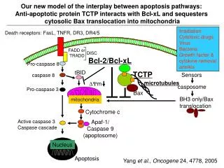
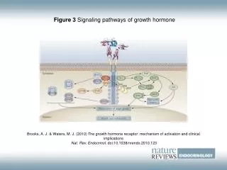
Simplified Signaling Pathways of Apoptosis Pollack M, Leeuwenburgh C: J Gerontol A Biol Sci Med Sci 2001;56:B475-482.
Caloric Restriction (CR) • CR increases mean and maximum life-span improves neuronal and muscle function as well as reduces cardiomyopathy. • 1) reduces oxidant production • 2) improves calcium homeostasis • 3) reduces inflammation • 4) reduces DNA damage
Mitochondrial Dysfunction and AgingDrew, B., Phaneuf, S., Dirks, A., Selman, C., Gredilla, R., Lezza, A., Barja, G., and Leeuwenburgh, C. (2003). Am J Physiol Regul Integr Comp Physiol 284, R474-R480. Leeuwenburgh, C., Wagner, P., Holloszy, J.O., Sohal, R.S., and Heinecke, J.W. 1997.. Arch Biochem Biophys 346:74-80. • Oxidative stress • mt-DNA damage • mt-DNA deletions • Oxidized proteins • Lipid peroxidation • Lipid-adduct formation • Decrease in repair systems
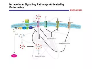
Endoplasmic (Sarcoplasmic) Reticulum “Stress” Damage due to reactive nitrogen and oxygen species Squier, T.C., and D.J. Bigelow. 2000. Protein oxidation and age-dependent alterations in calcium homeostasis. Front Biosci. 5:D504-26.
The effects of age and calorie restriction on TNF- / NF-B signaling in skeletal muscle Phillips and Leeuwenburgh (unpublished)
Myocardial Aging, Sarcopenia, Neurodegeneration, Oxidative Stress and Apoptosis • in total number of skeletal and heart myocytes as well as neurons with age • May lead to accelerated decline in cardiac functional capacity, sarcopenia, neurodegenerative diseases • Oxidative Stress and Apoptosis may be one major factor
Neuron loss in the brain with age and improved brain function with CR • Improved responses to enclosed alleys • Previous studies on neuronal loss with aging reported disparate results. • Studies often measured neuron density in a given structure instead of total neuron number. • Some studies suggest that most neocortical areas and certain hippocampal subfields lose 25 to 50% of their neurons with age in humans. • Specific regions of the brain appear to be effected (hippocampus, hilus of the dentate gyrus and the subiculum) Morrison, J. H., and Hof, P. R. (1997) Science 278, 412-419; Mattson, M. P. (2000). Nat Rev Mol Cell Biol 1, 120-129. Ingram, D. K., Weindruch, R., Spangler, E. L., Freeman, J. R., and Walford, R. L. (1987) J Gerontol 42, 78-81. Ingram, D. K., Chefer, S., Matochik, J., Moscrip, T. D., Weed, J., Roth, G. S., London, E. D., and Lane, M. A. (2001) Ann N Y Acad Sci 928, 316-326.
Is there skeletal muscle fiber loss with age in humans and animals? Animals Humans Bua EA, McKiernan SH, Wanagat J, McKenzie D, Aiken JM. Mitochondrial abnormalities are more frequent in muscles undergoing sarcopenia.J Appl Physiol. 2002 Jun;92(6):2617-24. Lexell J, Taylor CC, Sjostrom M. What is the cause of the ageing atrophy? Total number, size and proportion of different fiber types studied in whole vastus lateralis muscle from 15- to 83-year-old men. J Neurol Sci. 1988 Apr;84(2-3):275-94. Pesce V, Cormio A, Fracasso F, Vecchiet J, Felzani G, Lezza AM, Cantatore P, Gadaleta MN. Age-related mitochondrial genotypic and phenotypic alterations in human skeletal muscle Free Radic Biol Med,1;30(11):1223-33, 2001.
Muscle for control of urination 5-weeks 56-years 81-years
Myocyte Cell loss During Aging and CR Type II Fiber Deep portion of VL muscle is a type 2 fiber Type I Fiber Lee, C.M., Aspnes, L.E., Chung, S.S., Weindruch, R., and Aiken, J.M. 1998. Influences of caloric restriction on age-associated skeletal muscle fiber characteristics and mitochondrial changes in rats and mice. Ann N Y Acad Sci 854:182-191.
Muscle Function with Age and Calorie Restriction Type II Fiber Type I Fiber Anthony M. Payne, Stephen L. Dodd, and Christiaan Leeuwenburgh. Life-long calorie restriction in Fischer-344 rats attenuates age related loss in skeletal muscle specific force and extracellular space. J Appl Physiol (September 12, 2003). 10.1152/japplphysiol.00758.2003.
A – EDL, Young B – EDL, Old C – EDL, Old-CR D – Soleus, Young E – Soleus, Old F – Soleus, Old-CR Scale bar, 100 μm CR attennuated the age associated rise in extracellular space in the fast extensor digitorum longus (Type 2) muscle
Does caloric restriction attenuate age related alterations in apoptosis and cell signaling? • Are caspases effected? • Inhibitors of caspases? • Are other key pro- and anti-apoptotic proteins effected? • And do they influence the apoptotic potential (survival/death)?
Caspase-3; cleaved caspase-3; and X-linked inhibitor-of-apoptosis (XIAP) protein content as well as the enzymatic activity of caspase-3 in the gastrocnemius muscle
Oxidative damage, mtDNA deletions, and ETC abnormalities are co-localized along a single muscle fiber and exhibit atrophy Wanagat et al. 2001. Mitochondrial DNA deletion mutations colocalize with segmental electron transport system abnormalities, muscle fiber atrophy, fiber splitting, and oxidative damage in sarcopenia. Faseb J. 15:322-32 Bua EA, McKiernan SH, Wanagat J, McKenzie D, Aiken JM. Mitochondrial abnormalities are more frequent in muscles undergoing sarcopenia. J Appl Physiol. 2002 Jun;92(6):2617-24.
Fiber Splitting COX--/SDH++ Wanagat et al. 2001. Mitochondrial DNA deletion mutations colocalize with segmental electron transport system abnormalities, muscle fiber atrophy, fiber splitting, and oxidative damage in sarcopenia. Faseb J. 15:322-32
Koseki, T., Inohara, N., Chen, S., and Nunez, G. (1998) ARC, an inhibitor of apoptosis expressed in skeletal muscle and heart that interacts selectively with caspases. Proc Natl Acad Sci U S A 95, 5156-5160 Neuss, M., Monticone, R., Lundberg, M. S., Chesley, A. T., Fleck, E., and Crow, M. T. (2001) The apoptotic regulatory protein ARC (apoptosis repressor with caspase recruitment domain) prevents oxidant stress-mediated cell death by preserving mitochondrial function. J Biol Chem 276, 33915-33922
Cytosolic (A) and mitochondrial (B) ARC content in the gastrocnemius muscle
Coupling endoplasmic reticulum stress to the cell death program Rao, R.V., Castro-Obregon, S., Frankowski, H., Schuler, M., Stoka, V., del Rio, G., Bredesen, D.E., and Ellerby, H.M. 2002. Coupling endoplasmic reticulum stress to the cell death program: An Apaf-1-independent intrinsic pathway. JBC Nakagawa, T., and Yuan, J. (2000) Cross-talk between two cysteine protease families. Activation of caspase-12 by calpain in apoptosis. J Cell Biol 150, 887-894. Nakagawa, T., Zhu, H., Morishima, N., Li, E., Xu, J., Yankner, B. A., and Yuan, J. (2000) Caspase-12 mediates endoplasmic-reticulum-specific apoptosis and cytotoxicity by amyloid-beta. Nature 403, 98-103.
Caspase-12 and cleaved caspase-12 in the gastrocnemius muscle Nakagawa, T., and Yuan, J. (2000) Cross-talk between two cysteine protease families. Activation of caspase-12 by calpain in apoptosis. J Cell Biol 150, 887-894. Nakagawa, T., Zhu, H., Morishima, N., Li, E., Xu, J., Yankner, B. A., and Yuan, J. (2000) Caspase-12 mediates endoplasmic-reticulum-specific apoptosis and cytotoxicity by amyloid-beta. Nature 403, 98-103.
Caspase-12 • Intracellular calcium levels increase with age. Several studies have suggested that intracellular calcium handling is drastically improved following periods of caloric restriction • A ~350% increase in the expression of caspase-12 (caspase located at the sarcoplasmic reticulum) with age, CR reduced this age-associated rise • These data suggest that the caspase-12-mediated pathway of apoptosis may play a key role in sarcopenia and is attenuated by CR
Table 1. Overview of changes ( increase, decrease, no change) in apoptosis and apoptotic regulatory proteins in skeletal muscle with aging and calorie restriction (12AD v 26AD and 26CR v 26AD). Aging Calorie Restriction Gastrocnemius Muscle Markers for Apoptosis Apoptosis Caspase-3 (procaspase)1 Caspase-3 (cleaved caspase)1 Caspase-3 (activity)2 Mitochondrial Cytochrome c2 Apaf-11 Caspase-9 (procaspase)1 Caspase-9 (cleaved caspase)1 Apoptosis inducing factor1 Apoptosis inducing factor4 Inhibitors XIAP2 ARC1 ARC2 ARC3 Sarcoplasmic Reticulum Caspase-12 (procaspase)1 Caspase-12 (cleaved caspase)1 1Total tissue; 2Cytosolic; 3Mitochondrial; 4Nuclear. Dirks, A., and Leeuwenburgh, C. (2004). FRBM. Dirks, A., and Leeuwenburgh, C. (2002). Am J Physiol Regul Integr Comp Physiol 282, R519-527.
Table 2. Overview of changes ( increase, decrease, no change) in apoptosis and apoptotic regulatory proteins in cerebral cotices with age and calorie restriction (12AD v 26-27AD and 26-27CR v 27AD). Aging Calorie Restriction Markers for Apoptosis Apoptosis Cleaved PARP Caspase-3 (cleaved caspase)1 Caspase-3 (activity)2 Receptor Mediated Caspase-2 (cleaved caspase)1 Caspase-2 (activity)2 Mitochondrial Mediated Caspase-9 (cleaved caspase)1 Caspase-9 (activity)1 Cytochrome c2 Apaf-11 Inhibitors XIAP2 ARC2 1Total tissue; 2Cytosolic; 3Mitochondrial; 4Nuclear. Hiona, A., and Leeuwenburgh, C (2004). Unpublished data. Shelke, R. R. J., and Leeuwenburgh, C. (2003) Life-long calorie restriction (CR) increases expression of apoptosis repressor with a caspase recruitment domain (ARC) in the brain. FASEB J., 02-0803fje.
Summary • CR is able to attenuate the age-associated increase in apoptosis in skeletal muscle and neurons by altering several key apoptotic proteins towards cellular survival, thereby reducing the potential for sarcopenia and neurodegenerative diseases. • A diminished activation of mitochondrial-mediated, ER-mediated and death receptor-mediated pathways with life-long caloric restriction could have a profound affect on apoptosis and the susceptibility to apoptosis, cognition, as well as on muscle function. • The information obtained from these studies could potentially permit the development of physiological or genetic interventions that may attenuate the loss of skeletal muscle myocytes (sarcopenia) and neurodegeneration indicative of advancing age.
ACKNOWLEDGEMENTS Biochemistry of Aging Lab: Barry Drew, PhD (USA) Amie Dirks, PhD (USA) Sharon Phaneuf (USA) Suma K. PhD (India) Rajani Shelke, PhD (India) Colin Selman, PhD (Scotland) Tracey Philips, (Scotland) Mina Hiona (Greece) Young Yang (Korea) Collaborators: Gustavo Barja, PhD (Spain) Ricardo Gredilla (Spain) Steve Dodd, PhD (USA) Scott Powers, PhD (USA) Angela Lezza, PhD (Italy) Nicola Gadaleta, PhD (Italy) David Julian, PhD (USA) Tilman Grune, PhD (Germany) Funding Provided by: National Institute of Health National Institute on Aging Society of Geriatric Cardiology
Complete Data Web Page: http://grove.ufl.edu/~cleeuwen/