Download
1 / 38
440 likes | 650 Views
Cytoskeleton. Dr.rer.nat.,Dra. Asmarinah, MS. Depart. of Medical Biology Faculty of Medicine UI. Cytoskeleton. System of filaments in the cell, so that the cell have to be able to - change their shape move from place to place

E N D
Cytoskeleton Dr.rer.nat.,Dra. Asmarinah, MS. Depart. of Medical Biology Faculty of Medicine UI
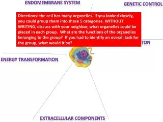
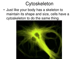
Cytoskeleton • System of filaments in the cell, so that the cell have to be able to • - change their shape • move from place to place • rearrange their internal components to grow, divide and adapt to changing circumstances • Examples of the cytoskeleton’s function: • Pulls the chromosoms apart at mitosis • Splits the dividing cell into two • Muscle contraction
Three types of cytoskeleton filaments: • Actin filaments = microfilaments • Determine the shape of the cell’s surface and necessary for whole-cell locomotion • 2. Microtubules • Determine the positions of membrane-enclosed organells and direct intracellular transport • 3. Intermediate filaments • Provide mechanical strength and resistance to shear stress
Each type of cytoskeletal filaments is constructed from smaller protein subunits • Microfilaments are made of subunits actin • Mictotubules are made of subunit tubulin • Intermediate filaments are made of smaller subunits, that elongated and fibrous All three types of cytoskeletal filaments are formed by assemblying (polimerisation) of their subunits through noncovalent linkages Nucleation is a step in the formation of a cytoskeletal polymer
The cytoskeleton and changes in cell shape A. Filament formation from a small protein subunits B. Rapid reorganization of the cytoskeleton in a cell in response to an external signal
The two ends of cytoskeleton filaments polymerize at different rates • plus end: the fast-growing end • minus end: the slow-growing end Within the cell, hundreds of different cytoskeleton-assosiated accessory protein regulate the distribution and dynamic behavior of the filaments, such as: - motor protein, to move or muscle contraction
The structure of an actin monomer and microfilaments • The actin monomer has a nucleotide bound in a deep cleft in the center of the molecule • Arrangement of monomer in a filament • Electron mikrographs of actin filaments Plus end upper cleft Minus end bottom cleft which bind ATP
Actin is a single globular polypeptide chain • Found in all eukaryote cell • Coded from many genes (polygene) • 90% amino acid homology in different spesies • three isoform: • α just in muscle sell • β present in all of the cell, excl muscle cell • γ ……………….”……………………………..
Actin-specific drug -Phalloidin: binds and stabilizes filaments -Cytochalasin: cap filaments plus end -Swinholide: severs filaments -Latrunculin: binds subunits & prevents their polymerization
A tubulin heterodimer is formed from α- and β-tubulin • monomer. The GTP molecule is tightly bound in α-tubulin, • but in β-tubulin is less tightly bound. • B. One tubulin subunit and one protofilaments are shown schematically • C. The microtubule is a stiff hollow tube formed from 13 protofilaments aligned in parallel • D. A short segment of a microtubule viewed in an electron microscope • E. Electron micrograph of a cross section of a microtubule showing a ring of 13 distinct protofilaments Plus end β-tubulin Minus end α-tubulin
Microtubule: • Found in eukaryote cell • There are + 6 isoforms of α-tubulin • + 6 isoforms of β-tubulin • coded by different gene • -Each of isoform has distinct function • -75% amino acid homology between yeast and human
Microtubule-specific drugs -Colchicine, colcemid: binds subunits and prevents polimerization -Vinblastine, vincristine: binds subunits and prevents polimerization -Nocodazole : binds subunits and prevents polimerization -Taxol: binds and stabilizes microtubules
A. Intermediate filaments monomer/subunit : a small filaments that are elongated and fibrous. B. Identical monomer form a dimer C. 2 dimers to form an antiparallel tetramer D. 2 tetramers packed together E. 8 tetramer twisted in a helixal array into ropelike filaments, which has 16 dimers in cross section
Intermediate filaments • Assembling and disassembling mechanismnot clear; might be protein phosphorilization disassambling
Motor protein (Molecular motor) • One of cytoskeleton-associate protein • Binds polarized filaments • Use ATP for the cell movement • Motor protein in cytoskeleton generate cytoskeleton sliding for • Muscle contraction • Cilia or flagella movement • Cell division
Three groups of cytoskeletal motor protein: • Myosin: associate with microfilaments for muscle contraction • Kinesin: motor protein which associate with mictotubule, play a role in chromosom separation in mitosis • Dynein: associate with microtubule, play a role in vesicle trafficking, reposisition of organell cell like mitochomdria as well as in cilia movement
Myosin (myosin II) 1 copi light chain myosin head Each of myosin head bind and hidrolisis ATP, to generate energy for microfilament movement to plus end direction
Electron micrograph of a myosin II thick filaments • Schematic diagram of myosin molecules which are aggregated by means of their tail regions with their head projecting to the outside of the filaments • A small section of a myosin II filaments as reconstructed from electron micrograph
Kinesin • is similar structurally to myosin • Most of them carry a binding site in the tail for either a membrane-enclosed organelle or another microtubule • Play a role in formation of spindel fiber in chromosom separation process in mitosis Dinein Largest of the known molecular motor and also fastest.Axonemal dynein can move microtubules at remarkable rate of 14 um/sec, the fastes kinesins can move their microtubules at about 2-3 um/sec
Flagella dan Cilia • Built from microtubules and dynein. The movement of them is produced by bending of its core, which is called axoneme. • The axoneme is composed by • 9 doublet microtubule • 1 pair microtubule in the central of axoneme • Dinein, consist of 2 kind of type: outer and inner • Another protein, such as nexin
Muscle contraction • Used for running, walking, swimming and flying in vertebrates • Depend on the ATP-driven sliding of highly organized arrays of actin filaments againts arrays of myosin Contractile elements of the muscle cell miofibril, is a cylindrical structure 1 – 2 um in diameter; consist of contractile units, called sarkomer
Skeletal muscle myofibrils A B C C • Electrone micrograph of longitudinal section through a skeletal muscle • Detail of skeletal muscle shown sarkomer • Schematic diagram of a single sarkomer
Organization of accessory protein sarkomer Titin: extends from Z disc to M Line, is closely associated with myosin. The rest of this protein is elastic Nebulin: coat actin filament Tropomodulin: cap the minus end of actin filament
Muscle Contraction Muscle contraction depends on the sliding of myosin and actin filaments, and is initiated by a sudden rise in cytosolic Ca2+ concentration The cycle of structural changes used by myosin to walk along an actin filament
Assesory proteins in muscle cell which are activated by increase of Ca2+ concentration : • Tropomiosin, associated with microfilaments • Troponin
Ca2+ + Troponin conformational changes Positional changes of tropomiosin Miosin head binds microfilaments Muscle contraction The Mechanism of muscle contraction
Sliding-filament model of muscle contraction. The actin (red) and myosin (green) filaments in a sarcomer slide past one another without shortening