Download
1 / 30
310 likes | 335 Views
Functional Hallux Limitus (FHL) is a condition affecting foot mechanics, leading to gait abnormalities and joint issues. Explore its origins, impact on gait mechanics, and progression to deformities in this detailed study.

E N D
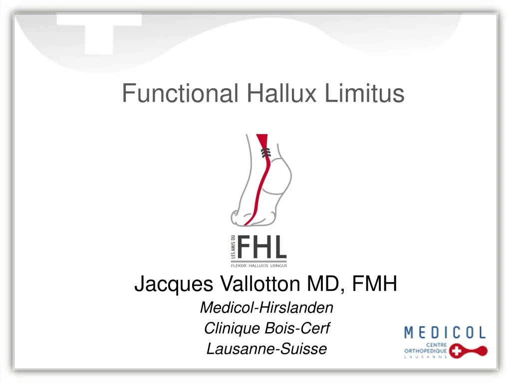
Functional Hallux Limitus Jacques Vallotton MD, FMH Medicol-Hirslanden Clinique Bois-Cerf Lausanne-Suisse
“Columbus Egg” of Orthopedics Functional Hallux Limitus (FHL) is a loss of MTP joint extension during terminal stance when the weight-bearing foot is in maximal dorsi- flexion and it constitutes a sagittal plain blockade. Substantial evidence highlight the tenodesis effect of the Flexor hallucis longus(Fhl) tendon during his passage in the retrotalar pulley as one of several origins of functional hallux limitus. Monument to the discovery of America by Columbus in the shape of an egg in Sant Antoni de Portmany, Ibiza, Spain
Topographic anatomy Conflict areas Origin Inferior 2/3 of posterior fibular surface
During the evolution • The hallux and its flexor hallucislongus tendon have evolved from a prehensile function to a propulsive one. • The muscle became more powerful • Talus has been anteriorised • The ankle plantarflexion decreased and the dorsiflexion increased.
Critical SituationThe retrotalar tunnel A B C 2 3 1 2 3 4 1 1 3 Subtalar Joint Subtalar Joint 2 Talus axial view 1.Postero-medial talar process 2. Retrotalar pulley 3. Postero-lateral talar process 4. Fhl tendon
Flexor Hallucis Longus This tenodesis effect • Derails the normal gait pattern • Desynchronizes the normal passage from pronation to supination and vice versa This functional derangement acts like a cascade • Modifies body balance • Disrupts joint mechanics from toe to head
Normal gait : synchronism in rotation Stance phase: The lower limb is globally oriented in internal rotation (femoral and tibial internal rotation and foot pronation) Push off phase: Transition to supination and external rotation
Autosupport mechanisms Hicks Windlass (1954) Bosjen- Moller Calcanocuboidal Joint stability (1979) Locked wedge effect i.e. compressive loading of osseous structures.
1st MTP Joint ROM and Self Stabilization • In 1954 in the Journal of Anatomy, JH Hicks published a series of papers collectively called the Mechanics of the Foot, I-IV • Within these, the Windlass Concept was introduced • Hallux dorsiflexion causes an unstoppable arch raising with simultaneous lower leg external rotation
Normal hallux dorsiflexion: key for the Windlass mechanism to work • Windlass mechanism (Hicks 1954): Toe acts as a winch pulling Fascia Plantaris and Flexor Hallucis Longus • Shortens distance between 1st MTT and calcaneus • Foot supination • External rotation of lower extremity on the subtalar joint. Functional Hallux limitus = Windlass BLOCKED
Functional hallux rigidus Dannanberg HJ, 1993 Induces a time-lag with a prolonged pronation at the push-off phase when the knee is extending as the limb approaches push off, the knee is unable to acquire tibial external rotation Medial collapse of the knee in stance
Pathologies related to Fhl • Hallux valgus et rigidus • Tendinopathies (peroneals, TFL, …) • Periostitis and stress fractures • Ankle and knee(ACL) injuries • Anterior knee pain syndrome (AKPS) • Sacro iliac and lumbar dysfunction • Piriformis syndrome • etc …
“Hallux Valgus and the First Metatarsal Arch Segment: A Theoretical Biomechanical Perspective” • 1st MT rotates around axis that is almost in horizontal plane • 2 sesamoid bones are located in intrinsic muscles underneath MT head • Hallux carries 40% of body weight at end of stance phase FHL Ward M. et al Physical Therapy Journal, 2010
Associated Findings What is the predisposing factor for all these findings??? Most common risk factor is overpronation which causes the 1st ray to be unstable and therefore hypermobile during gait Patients often walk with the feet laterally rotated and overpronated to compensate for lack of dorsiflexion and/or great toe extension during gait Collapse of medial longitudinal arch shifts the joint axis of the 1st MT from horizontal to vertical • Pronated foot • Decreased ankle dorsiflexion • Hallux limitus or rigidus
Sagittal plane blockade and FHLEffects on balance and gait • Negative effect on balance • Reduced mobility of subtalar joint • Centre of average instantaneous pressures abnormally displaced towards the rear of the foot • Cascade of compensatory mechanisms during gait required to overcome sagittal plane blockade: • Increased dorsal flexion of ankle, flexion of knee, hip, lumbar spine, cervical spine • The sagittal plane blockade increases the foot pronation in late stance • Medial collapse of foot, knee, hip
How The Pathology Progresses To Deformity • FHL progresses to either hallux rigidus or hallux valgus • Common characteristic decreased ability to dorsiflex the big toe joint when the heel comes off the ground in gait, forcing the joint to move when there is limited motion
Kelso SF, et al. Direction and range of motion of the first ray.J Am Pod Med Assoc 72:600-605, 1982. Why some people have HV and others HR? large range of motion of the first ray +highly tilted big toe joint axis + subluxation HV deformity smaller range of motion of the first ray + slight tilt of joint axis HR deformity
Clinical Examination High degree of suspicion Clinical Diagnosis • Typical callus formation • De-charging head of the 1st metatarsal • IP Hyperextension (occasionally)
A new clinical test • Specialized three phase test allows to differentiate the existence of Hallux Rigidus from the eg the pseudo tenodesis effect of the Fhl tendon at the posterior aspect of the subtalar joint.
B C A Clinical Examination FHL Stretch Test 1.Ankle in plantar flexion. Verify full ROM of the 1st MP joint. 2. Ankle in dorsiflexion by pressure applied at the metatarsal heads 3.Forced passive dorsiflexion of the MP1 joint. Inability to dorsiflex = Positive test
Gait analysis appui monopodal image podoscopique
B A Our experience Blue arrow: Fhl tendon. Red arrow: Retrotallar pulley. A: before resection. B: after resection More than 500 arthroscopic Fhl tendon tenolysis
The average pressure under the Hallux is statistically significantly lowered, postoperatively reflecting the repartition of the pressure between the Hallux and the augmented area under the first metatarsal head. The time of support in dynamic conditions under the head of the first metatarsal head did significantly change after surgery, which means that the foot becomes a more stable structure for balance and gait.
Conclusions • Based on our patient cohorts, FHL can be considered as a factor implicated in the pathogenesis of various lower limb pathologies • Careful examination is mandatory in order to reveal the pathognomonic symptoms usually masked especially in young adults. • Postural analysis has shown a remarkable postoperative restoration of the functional anatomy of the forefoot. • Postoperative clinical improvement, patient satisfaction and return to previous activities was good and excellent in more than 90%
Future steps • Define a clear gait pattern pathognomonic of FHL pathology. • Imaging modalities to further investigate the anatomical structure of pulley • Illustrate the correlation of FHL with other pathologies by use of larger populations • Implement 3D gait analysis technology to further delineate the implications of FHL in the context of functional anatomy and unity of form and movement