Download
1 / 80
830 likes | 1.74k Views
Learn about different types of spinal injuries, initial management principles, and neurological assessments. Understand the importance of careful handling to prevent further damage. Presented by Dr. Fadel Naim, an Orthopedic Surgeon.

E N D
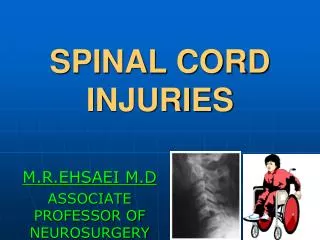
Spinal injuries Principles and treatment Presented By: FadelNaim M.D. Orthopedic Surgeon Faculty of Medicine IUG
Background • Approximately half the injuries occur in the thoracic, lumbar, and sacral areas • The other half occur in the cervical spine. • The average age at injury is 32 years • 55% of those injured are aged 16-30 years • Approximately 80% of patients are male
Spinal injuries can be described as: • Fractures • Fracture dislocations • SCIWORA / SCIWORET • Penetrating injuries
All injuries can stable or unstable • All patients with x-ray evidence of injury and all those with neurologic deficitsshould be considered to have an unstable spinal injury.
Blunt trauma • Motor vehicle accidents • Falls • Exlopsive injuries • Sport injuries
Spine injuries can result from • Axial loading • Flexion • Extension • Rotation • Lateral bending • Distraction
Spine Trauma suspect if • Head Trauma/ Loss of cosciousness • FFH • # calcaneum
Initial management • The goals for the emergency physician are to: • Establish the diagnosis • Initiate treatment • Prevent further neurologic injury from either: • pathologic motion of the injured vertebrae • secondary injury from the deleterious effects of cardiovascular instability • respiratory insufficiency.
Prehospitalcare: • stabilize and immobilize the spine on the basis of mechanism of injury, pain in the vertebral column, or neurologic symptoms. • Patients are usually transported to the ED with a cervical hard collar on a hard backboard.
Initial management • ABCs & Immobilization • Rigid collar/ backboard • Airway/ ventilatory support • Hemodynamic support
Beware • Excessive manipulation and inadequate immobilization of a patient with a spinal cord injury can cause additional neurological damage and worsen the patient’s outcome
Spine Trauma: Clinical : Exam • Look • Skin: Bruise,Wound • Posture • Feel • Tender • Swelling • Bony landmarks • Do NOT move !!
Initial management • Neurological exam: • performed as soon as the patient is hemodynamically stable: • Motor • Sensation • DTRs • digital rectal exam
If patient is conscious: radiographs and full exam • spinal injury can be excludes if: • No pain • Normal clinical examination of spine • Normal neuro exam • If the patient is unconscious : • spinal injury unless proven otherwise……? • neuro exam…radiographs… other urgent surgeries…
Initial Assessment Motor Examination • Upper extremity • C5-shoulder abduction • C6-wrist extension • C7-wrist flexion • C8-finger flexion • T1-finger abduction
Initial Assessment Motor Examination • Lower extremity • L1-hip flexion • L2-hip adduction • L3-knee extension • L4-ankle dorsiflexion • L5-toe extension
Initial Assessment • Dermatomes
Nerurogenic Shock vs Spinal Shock • Neurogenic shock results from impairment of the descending sympathetic pathways in the spinal cord resulting in loss of vasomotor tone and loss of sympathetic innervation to the heart • The result is: • Hypotension • Bradycardia
Nerurogenic Shock vs Spinal Shock • Spinal Shock results immediately after severe cord injury • a state of diminished excitability of the spinal cord.. • Due to sudden withdrawal of a facilitating or excitatory influence from the supraspinal centers. • Areflexic flaccid paralysis • Hypotension • Bradycardia • Duration varies: avg 3-4w
Clinical evaluation • Pain • Neurologic symptoms/signs • ASIA • Frankel scale • Image exams
American Spinal Injury Association (ASIA) • A = Complete – No Sacral Motor / Sensory • B = Incomplete – Sacral sensory sparing • C = Incomplete – Motor Sparing (<3) • D = Incomplete – Motor Sparing (>3) • E = Normal Motor & Sensory
Investigations: • Imaging the spine does not take precedence over life-saving diagnostic and therapeutic procedures
Image exams • Plain radiographs • Cheap and widely available • CT • 99% sensitivity for fracture • Quick • Disavantage – price and availability • MRI • Low specifity • Recommended to acces for injuries to soft tissue (ligamental, intervertebral disc, spinal cord injury) • Can distinguish betwen spinal cord edema and hemorrage • Very expensive • No better than CT as screening tool
Trauma series includes X-ray for : • lateral cervical • Chest • lateral thoracic • A/P and lateral lumbar • A/P pelvis
CT Scan • L3 unstable burst fracture
MRI Scan • Thoracic fracture subluxation with increased signal in conus medullaris
SCIWORA Spinal Cord Injury w/o Radiologic Abnormality • Spinal cord stretching leads to neuronal injury or even complete severing of the cord • Accounts for up to 70 % of ped. Spinal cord injuries • Most common in kids < 8 years • Paralysis may be present on arrival • Up to 30 % have a delayed onset of neurologic abnormalities • May not occur until up to 4-5 days after injury • Many have neurologic symptoms at the time of the injury, such as paresthesias or weakness, that have subsequently resolved
SCIWORETSpinal cord injury without radiographic evidence of trauma • First described in pediatric population (SCIWORA) • In adults, tends to affect the elderly • Much more prevalent in cervical spine as opposed to the thoracolumbar area. • Related to the degenerative changes in the c-spine
Classifications Necessary for…… • Uniform method of description • Directing treatment *** • Facilitating outcome analysis • Should be: Comprehensive Reproducible Usable Accurate
Fracture Classification • Fracture classification allows organization and treatment of fractures through protocols developed to maximize patient outcomes • Most classification schemes based on criteria for describing stability • The mechanism of trauma may be a more valuable parameter than fracture morphology for the classification and treatment
Anatomic Classification 2 or 3 Columns Denis ‘83 McAfee ‘83 Ferguson & Allen’84 Holdsworth’62 Kelley & Whitesides ’68
Mechanical Stability • 3-column theory(Denis ‘83) • middle = posterior ½ VB, posterior disc, post longitudinal lig • Disruption of 2/3 unstable • 2-column theory (Holdsworth,’53) • anterior= VB, disc, ALL, PLL • posterior= neural arch, Post lig complex
Anterior- Anterior longitudinal ligament, anterior half annulus fibrosus and vertebral body. Middle - Posterior longitudinal ligament, posterior half annulus fibrosus and vertebral body. Posterior - Osseous and ligamentous structures posterior to the posterior longitudinal ligament, (Interspinous ligaments)
Spine Fracture Types Compression fractures • Result from axial loading and flexion • Failure of the anterior column • Middle, posterior columns intact • Usually stable unless > 50% height • Unlikely to be directly responsible for neurologic damage
Burst Fractures • Axial load • Both anterior and middle columns fail • Retro-pulsion of bone and disk fragments into the canal • May cause spinal cord compression
Fracture Dislocations • Fracture-dislocations • Most damaging of injuries • Failure of all three columns • Compression, flexion, distraction, rotation, or shearing forces
Flexion- distraction • Seat belt–type injuries • Particularly where lap belts alone are used • Failure of both the posterior and middle columns • Intact anterior column prevents subluxation • Radiographic findings: • Increased height of the posterior vertebral body • Fracture of posterior wall of the vertebral body • Posterior opening of the disk space.
Whiplash injury • Sudden hyperextension and flexion • Increasing neck pain for the first 24hours • Associated headache, pain radiating to both shoulders and paraesthesia in hands • Reduced lateral flexion • Anterior longitudinal ligaments are torn causes dysphagia • Forward flexion against resistance is painful • 90% are asymptomatic after 2years • 10% still have pain
AP & lateral view radiographs of the lumbar spine demonstrates a narrowed T12 vertebral body height consistent with a compression fracture.
Lumbar spine fractures and dislocations. Plain radiographs reveal a fracture of L2 with L2-L3 subluxation. CT scan. Note the large amount of bone retropulsed inside the spinal canal
Scout view image from a spiral CT scan shows a complete subluxation fracture (curved blue lines) of the lower thoracic spine. Such an injury combines lateral displacement with rotational injury
Treatment Non-operative Treatment Surgical Treatment Rehabilitation
Goals of Non-operative Treatment • Preserve neurological function • Minimize deformity progression • Decrease pain • Restore Function ASSUMES THE SPINE IS STABLE