Download
1 / 53
530 likes | 909 Views
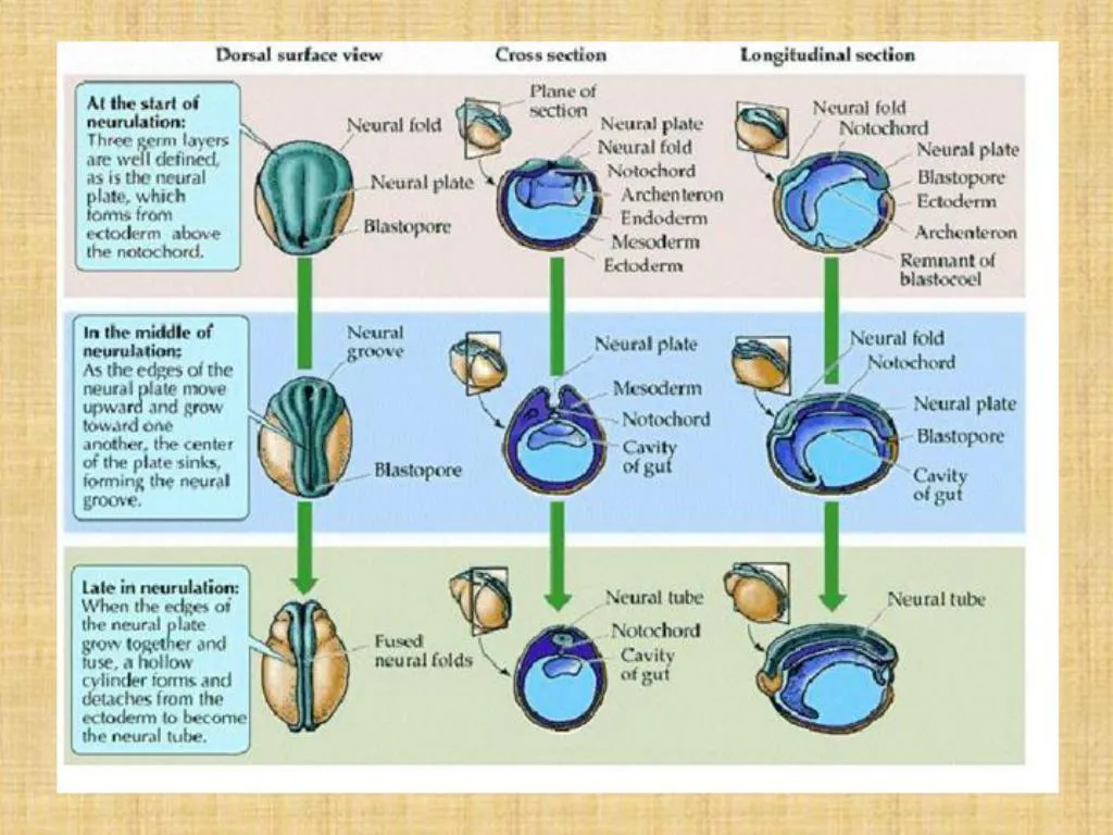
. . . . 24-hr Chick. 22-23 days pf Human. . 23-26 days pf Human. Formation of the Neural Tube. Secondary NeurulationOccurs beyond the caudal neuropore lumbar and tail regionExclusive mechanism for fish Starts with formation of medullary cordCavitation of cord to form hollow tube. Secondary Neurulation.

E N D
9. Formation of the Neural Tube Secondary Neurulation
Occurs beyond the caudal neuropore
lumbar and tail region
Exclusive mechanism for fish
Starts with formation of medullary cord
Cavitation of cord to form hollow tube
11. Differentiation of Neural Tube Major morphological changes: differentiation of brain vesicles and spinal cord
Differentiation of neural tube cells
Development of peripheral nervous system
13. Differentiation of Brain Vesicles Anterior neural tube bulges: 3 primary vesicles:
14. Then further differentiation into 5 secondary vesicles:
16. Differentiation of the Neural Tube Neural tube must maintain dorsal-ventral polarity
Sensory neurons- dorsal
Motor neurons- ventral
Accomplished by �inductive cascades�
Dorsal: BMPs from epidermis?Roof plate cells in neural tube?TGF-B cascade?Cell differentiation
Ventral: Sonic hedgehog from notochord and retinoic acid from somites?Floor plate cells of neural tube?shh gradient?Cell differentiation
18. Differentiation of the Neural Tube Histological changes
Neural tube initially a single layer of cells: germinal epithelium
Cells are called neural stem cells
Neurons
Glial Cells: Myelin sheath
28. Development of Peripheral Nervous System Divisions:
Autonomic NS
Sympathetic vs. Parasympathetic
Somatic NS
Anatomy
Sensory Neurons
Enter dorsal part of spinal cord
Soma located outside of cord?Dorsal ganglia
Form Dorsal Root of Spinal Nerve
Motor Neurons
Soma in ventral gray matter
Somatic NS: Neurons run directly from SC to muscle
Autonomic: 2 Neurons
Sympathetic: Ganglia near SC
Parasympathetic: Ganglia near or in or near organ
30. Development of Peripheral Nervous System Anatomy
Spinal Nerves:Spinal Cord
Sensory fibers of somatic nervous system: Dorsal root
Preganglionic neurons of sympathetic system: Ventral root
Motor fibers of somatic nervous system: Ventral root
Cranial Nerves: Brain stem
Sensory fibers of somatic nervous system: Dorsal root
Preganglionic neurons of parasympathetic system: Ventral root
Motor fibers of somatic nervous system: Ventral root
33. Origin of PNS Cells From neural tube:
All motor neurons of somatic nervous system
Preganglionic neurons of autonomic system
From neural crest:
Sensory nerves and associated ganglia
Postganglionic neurons of autonomic system
36. Neural Crest Cells Induced by organizing cells of notochord
Main functional groups:
Cranial neural crest:
Bones and connective tissue of face
Tooth primordia
Thymus, parathyroid, thyroid glands
Sensory cranial neurons
Parasympathetic ganglia and nerves
Parts of the heart (cardiac neural crest)
37. Neural Crest Cells Main functional groups:
Trunk neural crest:
Melanocytes
Sensory neurons
Sympathetic ganglia and nerves
Medulla of adrenal glands
40. Neural Crest Cells Migration:
Epithelial to mesenchyme transition
Migrational pathways are established by juxtacrine signals:
Fibronection, laminin in ECM + integrins
Ephrin proteins: Restrict movement
Contact inhibition
Use of existing structures
Migration ceases when these signals are reversed
42. Neural Crest Cells Differentiation:
Largely based on location along neural tube and their migration route:
43. Neural Crest Cells Differentiation:
Migration routes along trunk:
Ventral pathway: cells move through anterior portion of somite toward ventral side of embryo
Cells become: sensory neurons, sympathetic ganglia, medulla of adrenal gland
Dorsolateral pathway: cells move between epidermis and somite
Cells become: melanocytes
Basic organization of the PNS is established by the migratory pathways of the neural crest cells
45. Neural Crest Cells Differentiation:
How do they know what to become?
Most cells are pleuripotent- fate determined by position
Paracrine factors play a role
Example: Endothelin-3 and Wnt
Some exceptions: only NC cells from head make bone
Individual cells may differentiate early in migration
46. Differentiation of Neurons Within nerve tube:
Dorsal? Interneurons
Ventral? Motor neurons
47. Differentiation of Neurons Motor neurons:
Tissues they innervate depends on:
Anterior-posterior location along the nerve tube
When the cells were �born�
49. Axonal Pathways
50. Establishing pathways and connections:
Pathway selection
Target selection
Address selection
Axonal Pathways
51. Pathway Selection
Pathway axon takes influenced by extracellular matrix and cells encountered: signals by both paracrine and juxtacrine factors:
Cell adhesion and contact guidance: Haptotaxis
Growth cone repulsion
Ephrin and semaphorin proteins
Labeled Pathways Hypothesis: Pioneer Neurons
Diffusible molecules Axonal Pathways
52. Target Selection:
Neurotrophins on target cells (muscle or another neuron)
Address selection: constructing the synapse Axonal Pathways