Download
1 / 115
1.15k likes | 1.35k Views
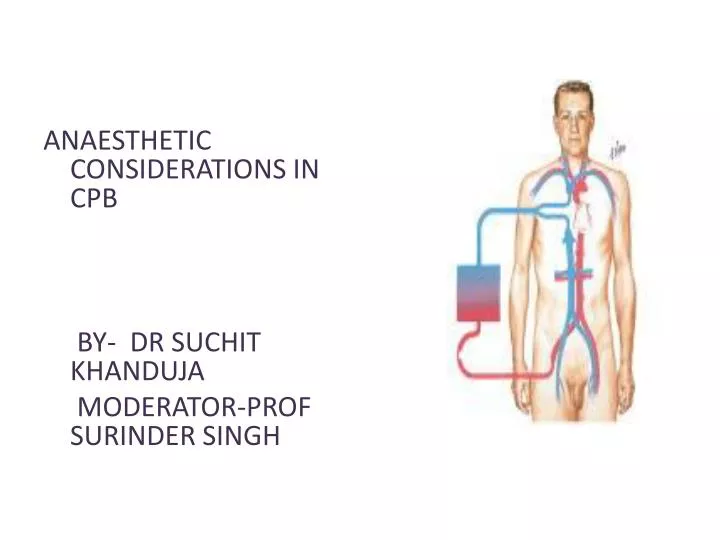
ANAESTHETIC CONSIDERATIONS IN CPB BY- DR SUCHIT KHANDUJA MODERATOR-PROF SURINDER SINGH . DEFINITION .

E N D
ANAESTHETIC CONSIDERATIONS IN CPB BY-DR SUCHIT KHANDUJA MODERATOR-PROF SURINDER SINGH
DEFINITION • “CPB is the technique whereby blood is totallyor partiallydiverted from the heart into a machine with the gas exchange capacity and subsequently returned to the arterial circulation at appropriate pressures & flow rates.”
HISTORICAL ASPECTS • Legllois (1812) : “circulation might be taken over for short periods” • Dr.John Gibbon(Philadelphia) 1953 : “performed ASD repair with the aid of CPB for the 1st time with the survival of patient.”
GOALS OF CPB To provide a still & Bloodless Heart with blood flow temporarily diverted to an Extracorporeal Circuit that functionally replaces the Heart & the Lung
GOALS OF CPB RESPIRATION Ventillation Oxygenation CIRCULATION TEMP. REGULATION (Hypothermia) Low blood flowso ed blood trauma ses Body Metabolism.
COMPONENTS OF CPB • TOTAL CPB : Systemic venous drainage CPB Circuit External oxygenator heat exchanger External pump arterial filterSystemic circulation. • PARTIAL CPB : Portion of systemic venous return (Rt. Heart) CPB .Undiverted blood Rt. Atrium Rt. Ventricle Pul. Circulation Lt. Atrium & Lt. Ventricle Systemic Circulation.
INTEGRAL COMPONENTS OF extracorporeal circuit • PUMPS • OXYGENATOR • Heat exchanger • Arterial filter • Cardioplegia delivery system • Aortic/atrial/vena cavalcannulae • Suction/vent
PATIENT ARTERIAL LINE FILTER RESERVOIR ROLLER PUMP OXYGENATOR HEAT EXCHANGER
ROLLER PUMP • Most commonly used. • Uses Volume displacement to create forward blood flow. • Non Pulsatile Blood Flow • By compressing Plastic Tubing b/w Roller & Backing Plate
Properly set occlusion causes minimal haemolysis • Occlusion is 100% in cardioplegia &vent pumps • Each pump indepedently controlled by a rheostat • Larger tubing and lesser rotations cause minimal haemolysis
Resistance= resistance of tubing+oxygenator+heatexcyhanger+filter+aorticcannulae+SVR • Usually line pr. depends on SVR and pump flow rate • Nl limit is 150-350 mm hg( >250 is seldom accepted)
DISADVANTAGE of producing PULSATILE FLOW • Bubble Formation • Damage to Blood Components. ADVANTAGE : • Improved Tissue Perfusion • Better Preservation of Organ Function (Brain , Kidney)
CENTRIFUGAL PUMP • Series of CONES that spin & propel blood forward by Centrifugal Force. • Safe • Reliable • Disposable • Simple to operate.
CENTRIFUGAL PUMP • ADVANTAGE • No back pressure when tubing is temporarily obstructed / kinked • Doesn’t produce spatulated emboli from compression of the tubing • Cannot pump large amt.of gas / gas emboli. • Less blood trauma • High vol. output with moderate pressures • DISADVANTAGE • Inability to generate pulsatile flow • Potential discrepancy b/w pump speed & actual flow generated.
Preferred over roller pumps in • Long-term CPB • In high-risk angioplasty patients • Ventricular assistance • Neonatal ECMO
Pressure-regulated pump • Operates under passive filling • After&pre-load sensitive • Pump-chamberofpolyurethane+peristalticpump • Not yet fully evaluated
OXYGENATOR • Where O2 & CO2 Exchange takes place. • Two Types : BUBBLE OXYGENATOR MEMBRANOUS OXYGENATOR
BUBBLE OXYGENATOR • Gas exchange by directly infusing the gas into a column of systemic venous blood. • A) OXYGENATING CHAMBERS:bubbles produced by ventilating gas through diffusion plate into venous blood column • CO2 bubble & oxygen plasma • Larger the No. of Bubbles ; Greater the efficiency of the oxygenator. • Larger bubbles improve removal of CO2 , diffuses 25 times more rapidly in plasma than O2 • Smaller bubbles are very efficient at oxygenation but poor in co2 removal
DEFOAMING CHAMBER • Defoaming of frothy blood. • Large surface area coated with silicone • This es the Surface Tension of the bubbles causing them to burst.
BUBBLE OXYGENATOR ADVANTAGE • Easy to assemble • Relatively small priming Volumes • Adequate oxygenating capacity • Lower cost. DISADVANTAGE • Micro emboli • Blood cell trauma • Destruction of plasma protein due to gas interface. • Excessive removal of CO2 • Defoaming capacity may get exhausted with time.
MEMBRANOUS OXYGENATOR • Gas exchange across a thin membrane • Eliminates the need for a bubble-blood contact & need for a defoamer; so more physiological. • Blood damage is minimum • Ideal for perfusions lasting for >2-3 hours. • 2 types of membrane: • SOLID:Silicone • MICROPOROUS:polypropylene,Teflon &polyacrylamide
MEMBRANOUS OXYGENATOR ADVANTAGE • Can deliver Air-O2 mixtures. • Hemolysis • Protein desaturation • Post-op bleeding • Better platelet preservation. DISADVANTAGE • Expensive • Large priming volume • Prolonged use pores may get blocked.
CIRCUITS • Drains Venous Blood by gravity into oxygenator & returns the oxygenated blood under pressure to the systemic circulation.
VENOUS DRAINAGE • Systemic venous blood (Rt.Heart)Oxygenator by • Direct Cannulation of SVC & IVC (BicavalCannulation) thru RA & joined to create a single drainage channel. • Single cannula into RA thru RA appendage. • Blood flow Oxygenator (Gravity) • Height Difference B/w Venacavae & Oxygenator > 20-30 cm.
Complications • arrhythmia • bleeding • ivc/svc tear • cannulamalposition • low return • inadequate height • malposition • kink,clamp,air lock
TUBINGS IN THE CIRCUIT • Non thrombogenic , Chemically Inert to prevent • clotting • Trauma to blood elements • Protein Denaturation • Smooth Internal Finish • Non Reactable Internal Surface • Durable to withstand high pressure & use of Roller pump
Made of • PVC • Polyurethane • Silicone • I.D . Ranges from 3/16- 5/8 inches • HEPARIN BONDED CIRCUITS ARE AVAILABLE
Disadvantages of plain circuits • Activation of platelets/coagulation factors • Post-op consumptive coagulopathyimmune reactions • More spallation Heparin coated circuits are • More hemo compatible • Cause less activation of platelets/white cells • Reduce heparin demand
INTRACARDIAC SUCTION • Blood will enter the heart • Coronary venous Return • Retrograde flow in AR. • Bronchial Arteries • CARDIOTOMY SUCTION • Spilled Heparinised Blood is Scavenged & returned back to patient. • Handheld Suckers are used to return this blood.
VENTRICULAR VENTING • LV Venting done to • Keep the operative field clear • Maintain Low LA & Pul.Venous Pressure • Remove air from Cardiac Chamber. •Blood from LV Reservoir Bag
RESERVOIR BAG • Collects the blood from VENOUS DRAINAGE & CARDIOTOMY SUCTION DRAIN PASSIVELY Reservoir Bag Oxygenator Heat exchanger Arterial Filter Patient. • Volume in the bag should not be allowed to empty to prevent massive emboli.
ARTERIAL RETURN • Ascending Aorta just proximal to Innominate Artery. • Femoral Artery in • Dissecting Aortic Aneurysm • For Reoperation • Emergency • Problems of Femoral Cannulation : • Sepsis • Formation of False Aneurysm • Development of Lymphatic Fistula.
ARTERIAL CANNULA • Is the Narrowest part of the circuit. • Should be as Short as possible. • As Large as the diameter of vessel permits.
Complications • Difficult Cannulation • Intramural Placement • Air embolism • Dislodgement of Cannula • Dissection • Arch Vessel Cannulation • Back Wall Injury
MICROPORE FILTERS: • Remove Particulate Matter (Bone , Tissue , Fat , Blood Clots etc.) • Pore Size : 30 – 40 • ULTRAFILTRATION : • Remove the excess fluid from the CPB.
PRIME FLUID • Ideally close to ECF. • Whole Blood NOT used : • Homologous Blood Syndrome. • Post Perfusion Bleeding Diathesis • Incompatibility Reactions. • Demand on Blood Banks. • Addition of Priming Fluid HEMODILUTION.
COMPOSITION OF PRIME : • Balanced salt soln. RL 1250 ml • Osmoticallyactive agent 100 ml (Mannitol, Dextran 40 , Hexastarch) • NaHCO3 50ml • KCl10ml • Heparin 1ml
PRIMING • Heme, nonheme • Decreases viscosity so better flow • Attenuates increased viscosity by hypothermia • Alters pharmacodynamics and kinetics of drugs • Decreases Hb but improves O2 delivery • Lowest acceptable value 8g/dl
Prediction of initial haematocrit during CPB Predicted Hct = Pt. RBC volume before CPB /Pt. EBV + CPB prime volume • EBV • Infants 80-85 ml/kg • Children 75ml/kg • Adult (male) 70ml/kg • Adult (female) 65ml/kg • 1U packed cells = 0.7 x 350 = 245ml • IU whole blood = 0.4 x 350 = 140ml
Amount of priming fluid • CVX CPCV = Pt. BV X PCV + PV X PCV • PT.BLOOD VOL. x PT. HEMATOCRIT = TARGET HCT X(PRIME VOL. + PT. BLOOD VOL.)
PATHOPHYSIOLOGY OF CPB • THREE MAJOR PHYSIOLOGICAL ABERRTIONS ARE: 1.LOSS OF PULSATILE FLOW 2.EXPOSURE OF BLOOD TO NON-PHYSIOLOGIC SURFACES & SHEAR STRESSES. 3.EXAGGERATED STRESS RESPONSE.