Download

1 / 12
130 likes | 249 Views
KERATOPLASTY VERSUS INTRASTROMAL SEGMENTS IN KERATOCONUS. HUMBERTO ESCORCE CURE CHIEF OF DEPARTMENT ANTERIOR SEGMENT INSTITUTO DE LA VISION - FUNDAVISIÓN BARRANQUILLA, COLOMBIA. THE AUTHORS HAVE NO COMMERCIAL INTEREST, NOR RECEIVE BENEFIT FROM, THE MATERIALS AND/OR PROCEDURES MENTIONED BELOW.

E N D
KERATOPLASTY VERSUS INTRASTROMAL SEGMENTS IN KERATOCONUS HUMBERTO ESCORCE CURE CHIEF OF DEPARTMENT ANTERIOR SEGMENTINSTITUTO DE LA VISION - FUNDAVISIÓNBARRANQUILLA, COLOMBIA THE AUTHORS HAVE NO COMMERCIAL INTEREST, NOR RECEIVE BENEFIT FROM, THE MATERIALS AND/OR PROCEDURES MENTIONED BELOW.
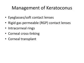
PURPOSE • Describe the topographic and visual acuity changes in a patient with a history of Keratoconus which performed a penetrating, keratoplasty and lasik in the left eye versus a treated with implant of intracorneal segments in the right eye.
METHODS • Descriptive study, clinical case, which discussed the case of a patient with diagnosis of bilateral Keratoconus, in whose left eye it was first performed a penetrating keratoplasty and one year later a Lasik . In the right eye were implanted Ferrara intracorneal segments. Were taken as the study variables topographic values pre surgical and postsurgical at 3 mm, 5 mm and 7 mm, visual acuity pre surgery and post surgery without and with correction with a 3-year follow-up.
RIGTH LEFT
COMPARISON OF TOPOGRAPHIC VALUES OF KERATOPLASTY AND INTRACORNEAL SEGMENTS FIRST YEAR
COMPARISON OF TOPOGRAPHIC VALUES OF KERATOPLASTY AND INTRACORNEAL SEGMENTS AT 3 YEARS
LEFT EYE RIGHT EYE
PREOPERATIVE POSTSURGlCAL
PREOPERATIVE POSTSURGlCAL
RESULTS • The preoperative uncorrected visual acuity was 20/400 logMAR (1.30) for both eyes. The preoperative best corrected visual acuity was 20/70 logMAR(0.54) for the right eye, and 20/80 logMAR (0.60) for the left eye. In right eye after intracorneal segment, corrected and uncorrected visual acuity was 20/30 logMAR (0.18). The left eye visual acuity was 20/80 logMAR (0.60) with and without correction posterior to keratoplasty. The lasik eye surgery is performed later obtained a visual acuity 20/50 logMAR (0.40) without correction, and 20/20 logMAR (0.00) with correction. • Topographicalchangesthatoccurred, in the right eye in which were placed intracorneal segments was observed greater stability of the topography and a reduction in keratometric values after three years, compared with the left eye who underwent keratoplasty, which presented an increase keratometric values after 3 years
REFERENCES • Intrastromalcorneal ring segments and corneal anterior stromal necrosis Jean-Louis Bourges, ThongThanTrong, Pierre Ellies, BenoitBriat, GillesRenardJournal of Cataract & RefractiveSurgeryJune 2003 (Vol. 29, Issue 6, Pages 1228-1230) • Ferrara intracorneal ring segmentsforkeratoconusSérgioKwitko, NórtonSouto SeveroJournal of Cataract & RefractiveSurgery - April 2004 (Vol. 30, Issue 4, Pages 812-820, DOI: 10.1016/j.jcrs.2003.12.005) • Ferrara intracorneal ring implantation and cataractsurgeryforthecorrection of pellucid marginal cornealdegenerationLeonardo Akaishi, Patrick F. Tzelikis, Irving M. RaberJournal of Cataract & RefractiveSurgery - November 2004 (Vol. 30, Issue 11, Pages 2427-2430, DOI: 10.1016/j.jcrs.2004.04.047) • Long-termfollow-up of intrastromalcorneal ring segments in keratoconusLeonardo Torquetti, Rodrigo FabriBerbel, Paulo FerraraJournal of Cataract & RefractiveSurgery - October 2009 (Vol. 35, Issue 10, Pages 1768-1773, DOI: 10.1016/j.jcrs.2009.05.036) • Keratitisafterintracorneal ring segmentinsertionforkeratoconusJames C. McAlister, NavidArdjomand, Luca Ilari, Lakhbir S. Mengher, David S. GartryJournal of Cataract & RefractiveSurgery - April 2006 (Vol. 32, Issue 4, Pages 676-678, DOI: 10.1016/j.jcrs.2005.09.026)