Download
1 / 18
220 likes | 570 Views
Donor lymphocyte infusion as a treatment of a complete DiGeorge syndrome. K.Zdráhalová 2 , E.Mejstříková 1,2 ,T.Kalina 1,2 ,P.Sedláček 2 , A. Janda 1 , H.Žižková 3 , Z.Sieglová 3 ,A.Šedivá 1 , J.Bartůňková 1 , J.Starý 2 , P.Kobylka 3 , P.Hubáček 2,4 , O.Hrušák 1,2

E N D
Donor lymphocyte infusion as a treatment of a complete DiGeorge syndrome K.Zdráhalová2, E.Mejstříková1,2,T.Kalina1,2,P.Sedláček2, A. Janda1, H.Žižková3, Z.Sieglová3,A.Šedivá1, J.Bartůňková1, J.Starý2, P.Kobylka3 , P.Hubáček 2,4, O.Hrušák1,2 2Department of Pediatric Hematology and Oncology, 1Department of Immunology, 3Institute of Hematology and Blood Transfusion, 4Department of Pediatrics, Prague, Czech Republic
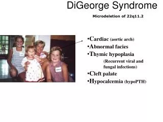
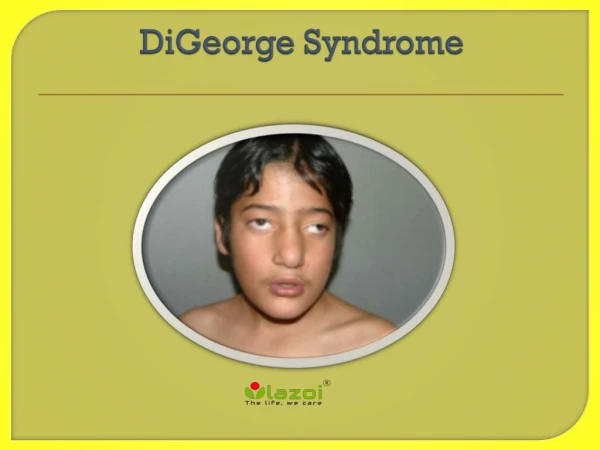
Introduction - DiGeorge syndrome • the most common deletion syndrome in humans – • monoallelic microdeletion of 22q11.2 (DiGeorge • and velo-cardio-facial sy, conotruncal anomaly) • variable phenotype even in pts with the same deletion • manifestation:("CATCH22") • mainly heart defects; hypoparathyroidism • thymic hypoplasia immunodeficiency, facialdysmorphism, developmental and behavioral problems • Deletion or interstitial deletion of 10p13 - other rare cause of DiGeorge sy (type II)
Patient: male, born June 18th 2004, now 10 months • Family history: healthy parents, 0 siblings • Personal history: 1st pregnancy, polyhydramnios amniocentesis normal karyotype 46, XY • term delivery, fetal hypoxia Cesarian section, • resuscitation, intubation, artificial ventilation • esophageal atresia + tracheoesophageal fistula: • D+2 operation • bilateral choanal atresia: D+5 operation • congenital heart defects: D+13 operation
Stigmatisation: • face • genitals • eyes • CNS • CHARGE association • (Coloboma, Heart disease, Atresia choanae, Retarded growth + • development and/or CNS anomalies, Genital anomalies and/or • hypogonadism, Ear anomalies and/or deafness) • Problems: • recurrent infections, septicemias • recurrent respiratory distress ventilation • apneas, irritability, states of altered consciousness
Immunology: • lymphocyte subsets in 2 months of age: • CD3+ 0% • NK 40% • CD4+ 0% • CD8+ 0% • CD19+ 58% • absent T cells • response to mitogens: absent • MRI -absent thymus at 12.8.04 prior DLI 16.12.05 10 CD4 (10^9/L) 1 CD8 (10^9/L) CD3 (10^9/L) CD19 (10^9/L) 0,1 NK (10^9/L) 0,01 0,001 0,0001
Complete DiGeorge Syndrome • (diagnosis at 2 months) • microdeletion 22q11 not found • prophylaxis started : • cotrimoxazole + itraconazole + IVIG
1st donor lymphocyte infusion age 6 months unrelated donor from register, 8/10 (B, Cw) no conditioning no GVHD prevention 1x 10⁶/kg CD3+; 0.2x 10⁶/kg CD34+ due to mistake non irradiated blood products administered (7 times prior 1st DLI, 1 time after 1st DLI)
1st donorlymphocyteinfusion(cont.) Between 1st and 2nd DLI • Complications:D+10: • isolated skin • aGVHD • (stage 3, grade II) • sepsis • cardiopulmonary • instability • capillary leak sy 10 after ATG 8 10 days post 1st DLI 0 1 CD3 (10^9/L) CD4 (10^9/L) 0,1 CD8 (10^9/L) 0,01 0,001 Chimerism: D+10 donordetected 0,0001 after ATG
Immunosupressive therapy: • rATG Fresenius(25mg/kg 3x D+10, D+12, D+14) • CsA • corticosteroids- MP (2mg/kg) • GVHD resolved • corticosteroids - 2 weeks 2 mg/kg, 1 week 1 mg/kg, • 1 week 0.5 mg/kg, then tapered (D+35) • CsA continued • ***** • D+33 last extubation! - aged 7 months
2nd donor lymphocyte infusion age 7 months, D+36 after 1st DLI the same donor no conditioning prevention of GVHD: CsA (continued) 0.89x 10⁶/kg CD3+
2nd donor lymphocyte infusion (cont.) EBV • Complications: • D+27EBV infection: • (B cell proliferation, • oligoclonality, IgM; • no clinical • manifestation) • withdrawal of CsA • Rituximab(375 mg/m2) • proliferation of • CD8+ activated T • cells started 10 rituximab CD3 CD8 10^9/L CD4 1 0 27 days post 2nd DLI 0,1 0,01 0,001 0 CD19
Chimerism after 1st and 2nd DLI chimerism in FACSorted T lymphocytes CD3+ (D+41 and D+55 CD4+ and CD8+): mainly donor chimerism in non separated blood: recipient mainly, donor detected proliferation of activated T cells, severe liver GVHD acute GVHD 1st DLI 2nd DLI d 0 d 8 d 19 d 10 d 25 d 10 d 34 d 41 d 55 last non irradiated trf EBV infection and prior rituximab No proof of engraftment of non irradiated blood transfusions
D+34:jaundice - bilirubin 4 mg/dL • dif.dg.: • hepatic GVHD • EBV lymphoproliferation(EBV in blood 0; in organs?) • hepatic infection- not found • ********* • D+352nd Rituximab(375 mg/m2) • D+41 corticosteroids(MP 1 mg/kg) hepatic aGVHD • neutropenia - granulo 215! • D+55 preventive ATB, antimycotics
D+45 - D+49: agranulocytosis (0 granulo) • corticosteroids(MP 2 mg/kg) • CsA • G-CSF5x • D+52: granulo 3000 • bilirubin 13.7 mg/dL • isolated liver GVHD stage 3, grade III
D+56: rATG Fresenius 1 dose, 25mg/kg • D+57: bilirubin 23.8 mg/dL • isolated liver GVHD stage 4, grade IV • ********* • corticosteroids (D+63 1.5 mg/kg, D+83 1 mg/kg) • CsA continues • Current status: • D+108 after 2nd DLI, age 10 months • bilirubin 7.6 mg/dL • CD8 activated T cells absolutely decreased • slight gradual psychomotor development
Patient aged 10 months Thank you.