Download

1 / 39
390 likes | 687 Views
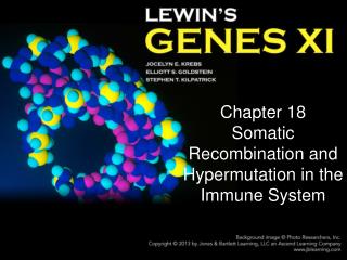
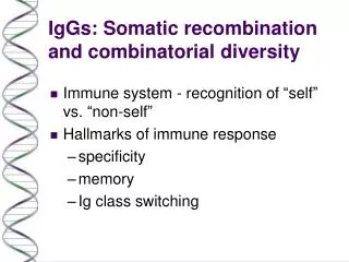
*. *. D J H rearrangement. V DJ H rearrangement. Primary RNA transcript. Splicing and polyA mRNA. Nascent polypeptide. Mature μ heavy chain protein. Somatic recombination at the Ig heavy chain locus. Germline DNA. *. Variable Region gene Variable Region exon.

E N D
* * D JH rearrangement V DJH rearrangement Primary RNA transcript Splicing and polyA mRNA Nascent polypeptide Mature μ heavy chain protein Somatic recombination at the Ig heavy chain locus Germline DNA * Variable Region gene Variable Region exon Gene Segments VH = Variable DH = Diversity JH = Joining JH = Joining ≠ J chain J chain is the “joining chain” for polymeric Ig IgM and IgA
The Major Histocompatibility Complex (MHC) Peter Burrows2011 4-6529 peterb@uab.edu • What it is • What it does • Required for antigen recognition by T lymphocytes
Learning objectives • To understand: • The differences between how T and B cells recognize antigen • The similarities and differences between MHC class I and class II expression and function • The consequences of MHC polymorphisms
Differences in antigen recognition by B and T lymphocytes • T cell antigen receptor • T cell receptor (TCR) • T cells • Transmembrane protein • Transmembrane protein on effector cells • CD4 Helper T Cells • CD8 Cytotoxic T Cells • B cell antigen receptor • Immunoglobulin (Ig) (BCR) • B cells • Transmembrane protein • Secreted by effector cells (Plasma cells)
Review of Differences in T and B Cells • B cells • Recognize native protein antigens in solution or on cell surfaces • Secreted antibody is effector molecule • Antibodies can operate at a distance
Review of Differences in T and B Cells • B cells • Recognize native protein antigens in solution or on cell surfaces • Secreted antibody is effector molecule • Antibodies can operate at a distance • T cells • Recognize peptides from degraded antigens • Peptides are displayed on cell surfaces in association with specialized proteins (MHC)
Antigen-antibody Interaction Heavy Chain Lysozyme Antigenic Determinant Epitope Light Chain
Proteases TCR #1 TCR #2 Antigen must be “processed” inside of cells to be recognized by T cells Lysozyme
T cells recognize processed (degraded) protein antigens plus MHC TCR 2
Phagocytosis Processing of external antigens
Processing of internal antigens Death Signal! Viral proteins
Review of Differences in T and B Cells • B cells • Recognize native protein antigens in solution or on cell surfaces • Secreted antibody is effector molecule • Antibodies can operate at a distance • T cells • Recognize peptides from degraded antigens • Peptides are displayed on cell surfaces in association with specialized proteins (MHC) • Antigen-specific T cell functions require direct cellcell interactions
Major Histocompatibility Complex • Discovered using inbred strains of mice and examining tumor immunity
Normal skin rejected Tumor rejected!
Major Histocompatibility Complex - MHC • Discovered using inbred strains of mice and examining tumor immunity • Were really studying transplantation immunology – histocompatibility antigens • MHC is the major histocompatibility antigen that needs to be matched for organ transplantation • A complex of linked genes encodes the MHC proteins • Normal function of MHC is to display peptide antigens (self AND non-self) to T cells
MHC • MHC I and MHC II • Expression and function • Structure • Genes and polymorphisms • MHC and T cell responses
MHC Class I • Protein expressed on all nucleated cells • Presents peptide to CD8 T cells • Cytotoxic “killer” cells • Kill virus infected cells Virus infected cell Tc killing virus infected cells
MHC Class II • Expressed by specialized antigen presenting cells (APC) • Dendritic cells • B cells • Macrophages • Presents peptide to CD4 T cells • “Helper” T cells • Help B cells proliferate, differentiate, isotype switch • Help activate macrophages to kill intracellular pathogens
B Cell #1 Class I Class II Selfpeptide Foreign peptide BCR
CD4 T Cell B Cell #2 B Cell #1 TCR Class I Class II Selfpeptide Foreign peptide
MHC Structure Class I Class II
Class I and Class II MHC Molecules • Membrane bound glycoproteins • Structurally very similar • Both have 4 domains • 2 membrane proximal domains • 2 membrane distal domains that form a peptide binding cleft
CD8 T Cell Virus infected cell
CD4 T Cell Antigen Presenting Cell (APC)
MHC Polymorphisms • Allelic variations in MHC genes • Concentrated in the peptide binding regions of Class I and Class II
MHC Polymorphisms • Allelic variations in MHC genes • Concentrated in the peptide binding regions of Class I and Class II • Population level • In an individual, all DP are identical, all B are identical, etc. No gene rearrangements with the MHC!
Consequences of MHC Polymorphism • Organ and tissue transplants are difficult • Polymorphic residues change the peptide binding specificity of the MHC • If all MHC were identical, pathogens might avoid immune detection by mutation to prevent MHC binding
Tc Tc Why have two antigen recognition systems?