Download
1 / 84
880 likes | 1.85k Views
CARDIOMYOPATHY. Athena Poppas, MD Associate Professor of Medicine, Brown Medical School Director, Echocardiography Laboratory Rhode Island Hospital. Cardiomyopathies. Definition: diseases of heart muscle 1980 WHO: unknown causes Not clinically relevant

E N D
CARDIOMYOPATHY Athena Poppas, MD Associate Professor of Medicine, Brown Medical School Director, Echocardiography Laboratory Rhode Island Hospital
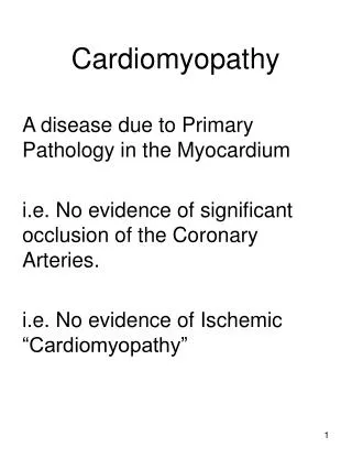
Cardiomyopathies Definition: diseases of heart muscle • 1980 WHO: unknown causes • Not clinically relevant • 1995 WHO: “diseases of the myocardium associated with cardiac dysfunction “ • pathophysiology • each with multiple etiologies
Cardiomyopathy • WHO Classification • anatomy & physiology of the LV • Dilated • Enlarged • Systolic dysfunction • Hypertrophic • Thickened • Diastolic dysfunction • Restrictive • Diastolic dysfunction • Arrhythmogenic RV dysplasia • Fibrofatty replacement • Unclassified • Fibroelastosis • LV noncompaction Circ 93:841, 1996
CM: Specific Etiologies • Ischemic • Valvular • Hypertensive • Inflammatory • Metabolic • Inherited • Toxic reactions • Peripartum Ischemic: thinned, scarred tissue
Dilated Cardiomyopathy • Dilation and impaired contraction of ventricles: • Reduced systolic function with or without heart failure • Characterized by myocyte damage • Multiple etiologies with similar resultant pathophysiology • Majority of cases are idiopathic • incidence of idiopathic dilated CM 5-8/100,000 • incidence likely higher due to mild, asymptomatic cases • 3X more prevalent among males and African-Americans
DCM: Etiology Ischemic Valvular Hypertensive Familial Idiopathic Inflammatory Infectious Viral – picornovirus, Cox B, CMV, HIV Ricketsial - Lyme Disease Parasitic - Chagas’ Disease, Toxoplasmosis Non-infectious Collagen Vascular Disease (SLE, RA) Peripartum Toxic Alcohol, Anthracyclins (adriamycin), Cocaine Metabolic Endocrine –thyroid dz, pheochromocytoma, DM, acromegaly, Nutritional Thiamine, selenium, carnitine Neuromuscular (Duchene’s Muscular Dystrophy--x-linked)
Prognosis depends on Etiology 1230 pts. referred for unexplained CM. Felker GM. NEJM 2000;342:1077
DCM: Infectious Acute viral myocarditis • Coxasackie B or echovirus • Self-limited infection in young people • Mechanism?: • Myocyte cell death and fibrosis • Immune mediated injury • BUT: • No change with immunosuppressive drugs
DCM: toxic Alcoholic cardiomyopathy • Chronic use • Reversible with abstinence • Mechanism?: • Myocyte cell death and fibrosis • Directly inhibits: • mitochondrial oxidative phosphorylation • Fatty acid oxidation
DCM: inherited Familial cardiomyopathy • 30% of ‘idiopathic’ • Inheritance patterns • Autosommal dom/rec, x-linked, mitochondrial • Associated phenotypes: • Skeletal muscle abn, neurologic, auditory • Mechanism: • Abnormalities in: • Energy production • Contractile force generation • Specific genes coding for: • Myosin, actin, dystophin…
DCM: Peripartum Diagnostic Criteria • 1 mo pre, 5 mos post • Echo: LV dysfunction • LVEF < 45% • LVEDD > 2.7 cm/m2 • Epidemiology/Etiology • 1:4000 women • JAMA 2000;283:1183 • Proposed mechanisms: • Inflammatory Cytokines: • TNFa, IL6, Fas/AP01 • JACC 2000 35(3):701.
PPCM: Prognosis • Death from CM: ’91-97 • 245 CM deaths in US, 0.88/100,000 live births, 70% peripartum • Increased risk with: • Maternal age • AA 6.4x greater • Whitehead SJ. ObGyn2003;102:1326. • Risk of recurrent pregnancy • Retrospective survey : 44 women (16 vs 28) • Reduced EF, CHF 44% vs 21%, mortality 0 vs. 19% • Elkyam U. NEJM.2001;344:1567. • DSE:contractile reserve reduced in patients • 7 women: change in Vcfc σES relationship • Lampert MB. AJOG.1997.176.189.
MECHANISMS IN HEART FAILURE Neurohormones Cytokines Oxidative stress Ischemic injury Myocardial disease Genetics Altered molecular expression Ultrastructural changes Myocyte hypertrophy Myocyte contractile dysfunction Apoptosis Fibroblast proliferation Collagen deposition Ventricular remodeling Hemodynamic Derangement Clinical Heart Failure Arrhythmia
Pathophysiology • Initial Compensation for impaired myocyte contractility: • Frank-Starling mechanism • Neurohumoral activation • intravascular volume • Eventual decompensation • ventricular remodeling • myocyte death/apoptosis • valvular regurgitation
Pathophysiology: Neurohumoral • Adrenergic nervous system • Renin-angiotensin-aldosterone axis • Vasopressin • Natriuretic peptides • Endothelin
Renin-Angiotensin-Aldosterone Pathways Angiotensinogen Renin ACE-inhibitor Angiotensin-I Chymase ACE Angiotensin-II Angiotensin receptor blocker Bradykinin degradation AT-1 Receptor Spironolactone Aldosterone
Vasoconstriction Aldosterone production Myocyte hypertrophy Fibroblast proliferation Collagen deposition Apoptosis Pro-thrombotic Pro-oxidant Adrenergic stimulation Endothelial dysfunction Angiotensin-II Effects
The Kidney in Heart Failure • Reduced renal blood flow • Reduced glomerular filtration rate • Increased renin production • Increased tubular sodium reabsorption • Increased free water retention (vasopressin)
Clinical Findings Biventricular Congestive Heart Failure -Low forward Cardiac Output -fatigue, lightheadedness, hypotension -Pulmonary Congestion -Dyspnea, -orthopnea, & PND -Systemic Congestion -Edema -Ascites -Weight gain
Physical Exam Decreased C.O. Tachycardia BP and pulse pressure cool extremities (vasoconstriction) Pulsus Alternans (end-stage) Pulmonary venous congestion: rales pleural effusions Cardiac: laterally displaced PMI S3 (acutely) mitral regurgitation murmur Systemic congestion JVD hepatosplenomegaly ascites peripheral edema
Diagnostic Studies CXR -enlarged cardiac silhouette, vascular redistribution interstitial edema, pleural effusions EKG –normal tachycardia, atrial and ventricular enlargement, LBBB, RBBB, Q-waves Blood Tests (ANA,RF, Fe2+, TFT’s,ferritin,) Echocardiography LV size, wall thickness function valve dz, pressures Cardiac Catheterization hemodynamics LVEF angiography Endomyocardial Biopsy
Influence of EF on Survival in Patients with Heart Failure Vasan RS et al. J Am Coll Cardiol. 1999;33:1948-55
Criteria for NYHA Functional Classification Class 1: No limitation of physical activity. Ordinary physical activity w/o fatigue, palpitation, or dyspnea. Class 2: Slight limitation of physical activity. Comfortable at rest, but symptoms w/ ordinary physical activity Class 3: Marked limitation of physical activity. Comfortable at rest, but less than ordinary activity causes fatigue, palpitation, or dyspnea. Class 4: Unable to carry out any physical activity without discomfort. Symptoms include cardiac insufficiency at rest. If any physical activity is undertaken, discomfort is increased. J Cardiac Failure 1999;5:357-382
Aim of Treatment • Preload reduction • Diuretics • venodilators • Vasodilators • ACEI • Inotropes • Acutely • Chronically • mortality
Vasodilator Agents in Heart Failure *Hydralazine and a long-nitrate shown to reduce mortality long-term # Other actions (aside from vasodilation) likely to be important
Electrical and Mechanical Ventricular Dyssynchrony • Experimentallyinduced LBBB has effect on: • expressionof regional stress kinases • calcium-handling proteins. • Expression of p38-MAPK(a stress kinase) is elevated in the endocardiumof the late-activated region, whereas phospholamban is decreased. • Sarcoplasmatic reticulum Ca2+-ATPaseis decreased in the region of early activation.
Deleterious Hemodynamic Effects of LV Dyssynchrony Diminished SV & CO due: • Reduced diastolic filling time1 • Weakened contractility 2 • Protracted MV regurgitation 2 • Post systolic regional contraction 3 Atrio-ventricular Intra-V Inter-V 1. Grines CL, Circulation 1989;79: 845-853 2. Xiao HB, Br Heart J 1991;66: 443-447 3. Søgaard P, JACC 2002;40:723–730 Cazeau, et al. PACE 2003; 26[Pt. II]: 137–143
CRT: Cardiac Resynchronization Therapy 1. Improved hemodynamics • Increased CO • Reduced LV filling pressures • Reduced sympathetic activity • Increased systolic function w/o MVO2 2. Reverse LV remodeling/architecture • Decreased LVES/ED volumes • Increased LVEF • Circ ’02, JACC ’02, JACC ’02, NEJM’02
Risk of Sudden Death c/w EF 1.00 1.00 0.98 0.98 p log-rank 0.002 0.96 0.96 0.94 0.94 Survival Survival 0.92 0.92 p log-rank 0.0001 0.90 0.90 0.88 0.88 A B Patients withoutLV Dysfunction (LVEF >35%) 0.86 0.86 0 30 60 90 120 150 180 0 30 60 90 120 150 180 Days Days Patients withLV Dysfunction (LVEF < 35%) No PVBs1-10 PVBs/h> 10 PVBs/h Maggioni AP. GISSI-2 TrialCirculation. 1993;87:312-322.
Women and Hypertension Prevalence of HTN in Women from NHANES-III. Burt VL. Hypertension ‘95
Diastolic Dysfunction • 40-50% of pts w/ CHF have nml LVEF • Vasan JACC ’99 • Grossman Circ ‘00 • Prevalence: • increases with age • higher in women • Etiology: HTN & LVH • Diagnosis: • MV& PV Doppler • TDI, Color m-mode
Echo Doppler Parameters Zile MR. Circ;105:1387
Diastolic Dysfunction Kawaguchi M. Circ 2003.107:714
Isolated Diastolic HF Isolated Systolic HF Systolic & Diastolic HF Zile MR. Circ;105:1387
What is the difference between systolic and diastolic LV dysfunction?
Hypertrophic Cardiomyopathy Left ventricular hypertrophy not due to pressure overload Hypertrpohy is variable in both severity and location: -asymmetric septal hypertrophy -symmetric (non-obstructive) -apical hypertrophy Vigorous systolic function, but impaired diastolic function impaired relaxation of ventricles elevated diastolic pressures prevalence as high as 1/500 in general population mortality in selected populations 4-6% (institutional) probably more favorable (1%)
Etiology Familial in ~ 55% of cases with autosomal dominant transmission Mutations in one of 4 genes encoding proteins of cardiac sarcomere account for majority of familial cases -MHC cardiac troponin T myosin binding protein C -tropomyosin Remainder are spontaneous mutations.