Download
1 / 28
1.74k likes | 6.51k Views
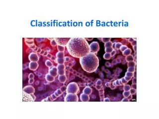
Classification of medically important bacteria. Bacteria. Unicellular, Microscopic, Prokaryotic Organisms, Multiply By Binary Fission. Comparison Between Bacteria And Fungi And Protozoa Bacteria Fungi & Protozoa Type Prokaryotic Eukaryotic Chromosome One Multiple

E N D
Bacteria Unicellular, Microscopic, Prokaryotic Organisms, Multiply By Binary Fission. Comparison Between Bacteria And Fungi And Protozoa BacteriaFungi & Protozoa Type Prokaryotic Eukaryotic Chromosome One Multiple (Number) Nuclear Absent Present Membrane
Comparison Between Bacteria and Fungi and Protozoa (Continued) BacteriaFungi & Protozoa Mitochondria Absent Present Ribosomes 70s 80s Sterols Absent (Except Usually In Mycoplasma) Present Cell Wall Rigid Layer Of No Peptido- Peptidoglycan Glycan (Absent In (In some cases Mycoplasma) cellulose present)
Bacteria can be divided into: • Filamentous Bacteria (Actinomycete) Most capable of branching • True (Euobacteria): Divide by Binary Fission • Spirocheates: Divide by Transverse Binary Fission • Mycoplasma Which Lack Rigid Cell Wall • Ricketssiae, and Chlamydia which are strict Intracellular parasites Vibrio (coma shape) Cocci Bacilli (rods)
Formal Rank Example Taxonomic Ranks ________________________________________________________________________________________________________________________ Kingdom Prokaryotae Division Gracilicutes Class Scotobacteria Order Eubacteriales Family Enterobacteriae GenusEschirichia, Streptococcus, Staphylococcus, Streptococcus, Klebsiella Species coli Pyogenes aureus pneumoniae pneumonia
The Gram stain, t bacteria into two main groups, is the first step in bacterial classfication & identification. • Bacteria stained purple are Gram + - their cell walls have thick petidoglycan and teichoic acid. • Bacteria stained pink are Gram – their cell walls have have thin peptidoglycan and lipopolysaccharides with noteichoic acid.
The Gram stain has four steps: • 1. crystal violet, the primary stain: followed by • 2.gramsiodine, which acts as a mordant by forming a crystal violet-iodine complex, then • 3. alcohol, which decolorizes, followed by • 4. safranin, the counterstain.
Is this gram stain positive or negative? Identify the bacteria.
Is this gram stain positive or negative? Identify the bacteria.
Gram staining tests the bacterial cell wall's ability to retain crystal violet dye during solvent treatment. Iodine is added as a mordant to form the crystal violet/iodine complex in order to render the dye impossible to remove. • Ethyl-alcohol solvent acts as a decolorizer and dissolves the lipid layer from gram-negative cells. This enhances leaching of the primary stain from the cells into the surrounding solvent. • Ethyl-alcohol will dehydrate the thicker gram-positive cell walls, closing the pores as the cell wall shrinks. • For this reason, the diffusion of the crystal violet-iodine staining is inhibited, so the bacteria remain stained.
Classification based on • Shape • Gram reaction • Oxygen • Free living & non free living
Simplified Classification of Medically – Important Gram-positive BacteriaFree living Arranged in Micrococcus Aerobes or clusters facultative Staphylococcus Anaerobes Cocci Arranged in Streptococcus chains Anaerobes Peptostreptococcus
Simplified Classification of Medically – Important Gram-positive bacteria Sporing Bacillus Aerobes or facultative anaerobes Corynebacterium Non- Listeria sporingLactobacillus Nocardia Mycobacterium RODS Sporing Clostridium Anaerobes Non- Actinomycosces sporing
Simplified Classification Of Medically – Important Gram-negative Bacteria Aerobes Neisseria Cocci Anaerobes Veillonella
Important Gram-negative Bacteria Aerobes Pseudomonas Salmonella Shigella enter Klebsiella obacProteus teriae Escherichia Facultative caeYersinia Anaerobes BACILLIrespirleigonellaBordetella Haemophilus zoonotBrucella Pasteurella francisella yersinia Vibrio(curved) Anaerobes Bacteroids Fusobacterium MicroaerophilicCamplylobacter
Simplified Classification Of Medically – Important Gram-negative Bacteria Aerobes Leptospira Spirochetes Treponema Anaerobes Borrelia Cell wall deficient bacteria------- Mycoplasma 2.Non- free living intra cellular— Rickettsia &chlamydia
Capsule Present in Certain Bacteria. • Polysaccharide; • occasionally protein e.g.Bacillus anthracis importance a. Inhibit Phagocytosis b. Antigenic
Microbiology And The Patient Medical Microbiology – concerned with: • Aetiology (cause) • Pathogenesis (Mechanism of production of disease) • Laboratory Diagnosis • Treatment of infection • Epidemiology (spread, distribution, prevalence of infection in the community) • Control and prevention in community
Laboratory Methods: Collection of specimens • Microscopy Stained Specimens Unstained Specimens • Culture • Identification of the organism • Tests for Antimicrobial agents serology • Demonstration of Abs
6) Understand the proper use of Clinical Lab. a) Specimen collection and handling b) Requesting appropriate tests c) Interpretation of results of Lab. tests • Correct selection, use, monitoring of anti-microbial therapy • Understand methods of prevention of infection e.g.Vaccine, chemoprophylaxis, hygiene, isolation etc.