Download
1 / 52
520 likes | 617 Views
SKELETAL SYSTEM. SKELETAL SYSTEM FUNCTIONS. Support (Primary function) Movement (Passive) Protection of Vital Organs Mineral Storage Blood Cell Formation (Hematopoiesis or Hemopoiesis). OSSEOUS C.T. Compact (dense) Bone Hard & heavy Forms surface & diaphysis Osteons = building blocks

E N D
SKELETAL SYSTEM FUNCTIONS • Support (Primary function) • Movement (Passive) • Protection of Vital Organs • Mineral Storage • Blood Cell Formation (Hematopoiesis or Hemopoiesis)
OSSEOUS C.T. • Compact (dense) Bone • Hard & heavy • Forms surface & diaphysis • Osteons = building blocks • Cancellous (spongy) Bone • Lightweight • Fills epiphyses, Contains red marrow • Trabeculae = building blocks • Matrix • Mineral Salts (hardness) • Collagen (strong & flexible)
Bone Cells • Osteoblasts – Secrete to form bone • Osteocytes • Mature bone cells • “Trapped” osteoblasts • Osteoclasts – destroy bone • Enzymes digest protein • Acids dissolve minerals • Forms Marrow Cavity; Involved in Remodeling
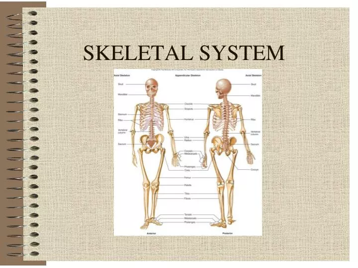
SKELETAL DIVISIONS • Axial • Appendicular
Classification: Shape & Location • Long • Short • Flat • Irregular • Sesamoid (develop in tendons; patella) • Sutural (between cranial bones)
LONG BONE ANATOMY • Diaphysis = shaft made of compact bone • Epiphyses = ends filled with spongy bone containing red marrow • Articular cartilage covers epiphyses • Epiphyseal line indicates earlier location of epiphyseal (growth) plate
LONG BONE ANATOMY • Periosteum is C.T. covering bone • Nutrient Foramina – holes allowing for penetration of arteries • Medullary cavity contains yellow marrow • Endosteum is C.T. lining medullary cavity
BONE DEVELOPMENT • Ossification = replacement of other connective tissue with bone • Begins during the 2nd month of gestation • Size increases until late teens (females) to mid-twenties (males) • Ossification processes include: • Intramembranous bone formation • Endochondral bone formation
INTRAMEMBRANOUS OSSIFICATION • Occurs in flat bones of skull, clavicles, mandible • Begins with fibrous C.T. membrane • Membrane calcifies & ossifies into bone • Fontanels • “Soft spot”, not yet ossified • Allows for birth & brain growth
Process of Intramembranous Ossification • Embryonic C.T. cells cluster & differentiate • Osteoblasts form & produce matrix = ossification center • Newly formed matrix calcifies • Osteocytes form
Process of Intramembranous Ossification • Trabeculae form; periosteum forms from surrounding condensed embryonic C.T. • Surface trabeculae fill with matrix, forming compact bone
ENDOCHONDRAL OSSIFICATION • Occurs in remainder of skeleton • Begins with hyaline cartilage model • Cartilage is replaced by bony tissue • Steps include: • Bone collar forms • Cartilage in shaft calcifies • Primary Ossification center forms in shaft • Secondary Ossification centers in epiphyses
APPOSITIONAL GROWTH • Bone Widens • Osteoclasts enlarge medullary cavity • Osteoblasts add bone to surface of diaphysis
BONE REMODELING • Replacement of old bone with new bone • Involves resorption (osteoclasts) & deposition (osteoblasts) • Alters bone shape in response to stress • Benefits: • Allows for growth • Removes injured bone • New bone is more resistant to fracture
FRACTURES AND THEIR REPAIR • Definition: Any break in a bone • Repair may take months • Requires: • Adequate minerals • Vitamins • Hormones • Weight-bearing exercise
STEPS IN FRACTURE REPAIR • Broken blood vessels form a hematoma (blood clot) • C.T. and Capillaries invade site; C.T. cells form fibrocartilage callus • Bony callus of spongy bone replaces fibrocartilage callus • Bony callus is remodeled
Types of Fractures • Open (Compound) – Broken bone ends protrude through the skin • Closed (Simple) – Bone does not penetrate the skin
Types of Fractures • Comminuted – splintered, crushed; small pieces between broken ends. Elderly. Most difficult to treat. • Greenstick – Partial fracture; one side breaks, other side bends. Children.
Types of Fractures • Impacted – One end of fractured bone forcefully driven into other end • Spiral – fracture spirals around long bone axis from twisting force
Types of Fractures • Pott’s – distal end of fibula, tibia or both • Colles’ – distal end of radius
Types of Fractures • Stress Fracture • Fracture without visible break • Microscopic fissures • No apparent damage to surrounding tissues • Result from repeated, strenuous activities • Can result from reduced calcium deposition in disease • 25% involve tibia
BONES AS LEVERS • Lever: A rigid rod that moves about a fixed point • Fulcrum: The fixed point around which a lever moves (joints) • Forces: Act to move levers at two points • Resistance: Force to be overcome • Effort or Work: Force required to overcome resistance; supplied by skeletal muscles
CLASSES OF LEVERS • First Class: The fulcrum is between the effort/force and the resistance • Seesaw • Tilting head backward
R R R R R R R F E E E E E E E FIRST CLASS LEVER
CLASSES OF LEVERS CONTINUED • Second Class: Resistance is between the fulcrum and the effort/force • Wheelbarrow • Rising up on one’s toes
R R R R R R R R F E E E E E E E E SECOND CLASS LEVER
CLASSES OF LEVERS CONTINUED • Third Class: The effort/force is between the fulcrum and the resistance • Most common type in the human body • Flexing the elbow
R R R R R R R R F E E E E E E E E THIRD CLASS LEVER
CLASSIFICATION OF JOINTS • Structural • Based on what tissues or structures are found between the bones • Fibrous, Cartilagenous, Synovial • Functional • Based on amount of movement (& amount of movement is determined by structures found between bones) • Freely movable, Slightly movable, Immovable
Pubic symphysis Functional: AmphiarthrosisStructural: Cartilagenous Knee Functional: DiarthrosisStructural: Synovial Sutures Functional: SynarthrosisStructural: Fibrous ARTICULATIONS: EXAMPLES
STRUCTURE OF A SYNOVIAL JOINT • Articular cartilage – covers bone ends • Synovial membrane – lines joint capsule • Synovial fluid – lubricates & nourishes cartilage • Synovial cavity – space between the bones • Joint capsule – fibrous C.T. • Ligaments – reinforce joint • Bursae – synovial sacs at other sites of friction