Download
1 / 51
820 likes | 2.15k Views
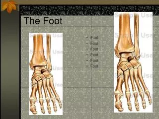
The Foot. Chapter 17. Foot Anatomy. 26 Bones 7 Tarsal 5 Metatarsal 14 Phalanges 38 Joints 4 Arches. Bones of the Foot. Bones of the Foot. Bones of the Foot. Tarsal Bones. Talus Calcaneus Navicular Cuboid Cuniforms Medial Intermediate Lateral. Metatarsals & Phalanges.

E N D
The Foot Chapter 17
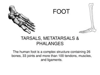
Foot Anatomy • 26 Bones • 7 Tarsal • 5 Metatarsal • 14 Phalanges • 38 Joints • 4 Arches
Tarsal Bones • Talus • Calcaneus • Navicular • Cuboid • Cuniforms • Medial • Intermediate • Lateral
Sesamoid Bones • 2 (medial and lateral) • Under great toe • Functionpulley, increase leverage of tendons that control great toe
Joints of the Foot • Tibiotalar • Talocrural • Subtalar • Talonavicular • Calcaneocubiod • Metarsocunieform
Joints of the Foot • Metatarsophalangeal Joint • Proximal Interphalangeal Joint • Distal Interphalangeal Joint
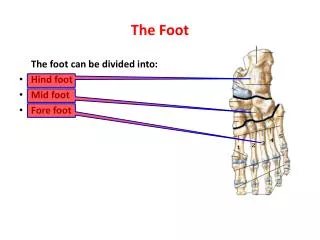
Regions of Foot • Forefoot • Metatarsals • Phalanges • Midfoot • Navicular • Cuboid • 3 Cuniforms • Hindfoot • Calcaneus • Talus
Plantar Fascia • Thick white band of fibrous tissue originating from the medial tuberosity of the calcaneus and ending at the proximal heads of the metatarsals • Work with ligaments to support arches during weigh bearing and downward forces
Foot Arches • Functions • Support body weigh in an economical fashion • Absorb the impact of walking, running, jumping or any other weight bearing activity • Provide a space on the plantar aspect of foot for blood vessels, nerves, and muscles
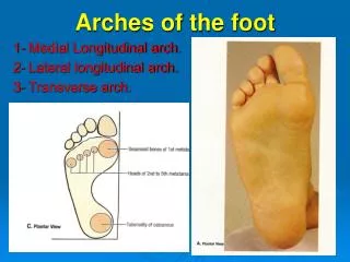
Medial Longitudinal Arch • Highest of 3 arches of foot • Calcaneus, Talus, Navicular, Cuniforms & 1st three metatarsals • Supports— • Ligaments: • Spring ligament • Plantar fascia • Tendons: • Tibialis posterior • Tibialis anterior
Lateral Longitudinal Arch • Lower and flatter • Calcaneus, Talus, Cuboid, 4th & 5th metatarsals • Supports— • Ligaments: • Short plantar ligament • Plantar fascia • Tendons: • Peroneus longus
Transverse Arch • Cuniforms, Cuboid, & 5th metatarsal
Shoe Wear Patterns • Excessive Pronation • Wear out front of shoe under 2nd metatarsal • Excessive Supination • Wear out lateral border of shoe • Common Misconception • Wearing out the back lateral corner of the shoe means you pronate • This is normal wear pattern
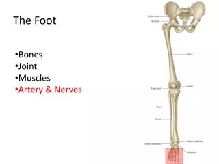
Posterior Tibial Artery Medial Malleolous Dorsalis Pedis artery Extensor Tendon Great Toe Pulse
Dorsiflexion Plantar Flexion Pronation Inversion Eversion Supination Foot Movements
Muscles of Foot Intrinsic Muscles Extrinsic Muscles Muscle outside a body part, organ, or bone Gastrocnemius • Relate to specific body part or bone • Flexor hallucis longus • Flexor hallucis brevis • Flexor digitorum longus • Extensor digitorum longus • Abductor hallucis • Abductor digiti minimi • Tibialis posterior
Fractures & Stress Fractures • Impair ability to perform competitively • NWB • More swelling & pain than ligament sprain • Point tenderness present • Obvious deformity often present • Usually occur acutely; result of traumatic episode
Jones Fracture • Fracture to the diaphysis at the base of the 5th metatarsal • Repetitive stress, direct force, or inversion and PF of foot • Healing slow; high nonunion rate
Retrocalcaneal Bursitis • Swelling of the bursa at the back of the calcaneus under the Achilles tendon • S/sxs: • Pain in heel • Painful to touch • Pain worse when rising on toes • Red, warm skin over back of heel
Plantar Fascia • Wide, non-elastic ligamentous tissue that extends from the anterior portion of calcaneus to heads of metatarsals • Supplies support to longitudinal arch
Plantar Fasciitis • Strain/irritation of the plantar fascia • Caused by: • Overuse • Unsupportive footwear • Tight Achilles tendon • Running on hard surfaces • Chronic irritation • Pain, tenderness on bottom of foot near heal (especially in am) • Untreated will lead to: • Bone imbalance • Heel spurs • Muscle strains • Shin splints
Plantar Fasciitis—Treatment • Correct training errors • Ice • Massage • Evaluate shoes & activity level • Arch support • Heel cup or cushion
Pes Planus • Flat foot • Associated with excessive pronation • Multiple causes: • Lack of shoe support • Weak muscles • Pain & weakness in medial longitudinal arch • Calcaneal eversion • Navicular bulging • Flattening of arch
Pes Cavus • aka Clawfoot, hollow foot • Associated with excessive supination • Shock absorption poor • General foot pain and metatarsalgia common • Abnormally short Achilles tendon • Calluses ball and heel
Arch Sprains • Ligaments stretch, thus fail to hold bones of foot in position • When arch weakened, it cannot absorb shock normally • Causes: • Overuse • Overweight • Fatigue • Training on hard surfaces • Non-supportive shoes • Shoes in poor condition
Turf Toe • Great toe strain • Hyperextension of the first MTP joint of the big toe • Treatment: • RICE & Support • Limit movement • Turf toe taping
Heel Spur • Bony growth on calcaneus • Causes painful inflammation • Aggravated by exercise • As foot flattens, plantar fascia is stretched & pulled where it attaches to calcaneus calcaneus reacts by forming spur of bony material
Heel Contusion • Irritation of the lateral aspect of the heel • Sudden stop-and-go or sudden change in movement
Heel Contusion—Treatment • Cold application before activity • Ice & elevation after activity • Absorb shock— • Heel cups • Donut pad
Sever’s Disease • Traction injury at the apophysis of the calcaneus where the Achilles tendon attaches • Young, physically active athletes • Comparable to Osgood-Shlatter’s disease (at tibial tubercle of knee) • Pain occurs during vigorous activity and does not continue during rest
Blisters • Occur on any part of body where there is friction • Most common on feet or heels • Treatment Goals: • Relieve pain • Keep from enlarging • Avoid infection
Blisters—Treatment • Wash area thoroughly • Use sterile blade to cut small hole in blister • Squeeze out clear fluid • Do not remove skin • Prevention: • Wear work gloves • Break in new skin • Petroleum jelly/skin lube • Adhesive bandage
Prevention of Foot Injuries • Selecting appropriate footwear • Using shoe orthotic • Foot hygiene
Rehabilitation of the Foot • Towel pulls • TheraBand® • Marble pick-up
Muscle Movement Foldable • Label each flap on of the directional terms • Toe Flexion • Toe Extension • Ankle Eversion • Ankle Dorsiflexion • Ankle Plantarflexion • Glue picture of movement on the under side of the flap. Write the muscle names under the correct flap. • Flexor Hallucis Longus • Extensor Digitorum Longus • Tibialis Posterior • Peroneus Longus • Extensor Hallucis Longus • Flexor Hallucis Brevis • Gastrocnemius • Peroneus Brevis • Soleus • Tibialis Anterior • Peroneus Tertius • Flexor Digitorum Longus