Download
1 / 50
540 likes | 801 Views
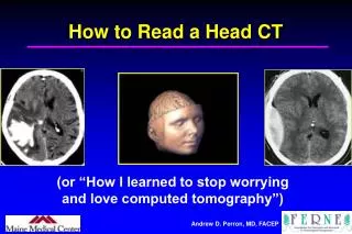
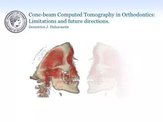
Applications of Magnetic Resonance Imaging (MRI) and Computed Tomography CT). Lecture 1 F33AB5. What are CT and MRI?. CT uses X-rays to produce tomographs (images of slices) MRI uses magnetic fields to probe the intrinsic magnetisation of hydrogen nuclei.

E N D
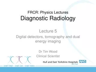
Applications of Magnetic Resonance Imaging (MRI) and Computed Tomography CT) Lecture 1 F33AB5
What are CT and MRI? • CT uses X-rays to produce tomographs (images of slices) • MRI uses magnetic fields to probe the intrinsic magnetisation of hydrogen nuclei http://www.mri-ny.com/scannersound.html
Overview • Advantages and problems of each technique • Anatomical imaging • Functional imaging Phillips
Problems of CT • Dose (fluroscopy/dynamic mode not possible) • (Speed- improving) • (3D- now available using helical scanning) • Artefacts behind bone
Advantages of CT • (Limited) soft tissue contrast • Spatial measurements exact (if set up correctly)
Problems of MRI • Not for people who are claustrophobic • Not for people with metal in their bodies • Susceptibility differences (eg between air and tissue) cause distortions in most sequences, compromising surgical planning • Can be slow (not EPI), can have motion artefacts • Can be expensive (£750k)
Advantages of MRI • Excellent (and controllable) soft tissue contrast • Much functional information • Steerable imaging planes • Safe • Hugely versatile
Anatomical- CT • Intracranial bleeds • Radiotherapy planning • low geometric distortion • CT contrast relates to radiation attenuation • Stereotactic surgery • low geometric distortion • Angiography
Anatomical- CT Chronic subdural haematoma • Intracranial bleeds • Radiotherapy planning • low geometric distortion • CT contrast relates to radiation attenuation • Stereotactic surgery • low geometric distortion • Angiography http://www.radiology.co.uk/xrayfile/xray/tutors/cttrauma/tutor.htm
Anatomical- CT • Intracranial bleeds • Radiotherapy planning • low geometric distortion • CT contrast relates to radiation attenuation • Stereotactic surgery • low geometric distortion • Angiography
Anatomical- CT Radiotherapy planning Real Time Multi-Trial Window http://www.adaclabs.com/prodSolu/rtp/3dtp/3dtp.shtml
Anatomical- CT Radiotherapy planning Dose distribution along path shown as histogram colored according to the volumes of interest. http://www.uke.uni-hamburg.de/institute/imdm/idv/publikationen/car1993/
Anatomical- CT • Intracranial bleeds • Radiotherapy planning • low geometric distortion • CT contrast relates to radiation attenuation • Stereotactic surgery • low geometric distortion • Angiography
Anatomical- CT MRI CT Brain with a deep central tumour CT generally has better geometric accuracy Patient a metal sterotactic frame, ( 'spots' around the head in the images). Streaking artifacts on the CT scans, because of beam-hardening effects. Dr Paul Morgan, from Academic Radiology
Anatomical- CT • Intracranial bleeds • Radiotherapy planning • low geometric distortion • CT contrast relates to radiation attenuation • Stereotactic surgery • low geometric distortion • Angiography
Anatomical- CT Angiography Left carotid artery showing aneurysm
Anatomical- CT Angiography Ascending aortic aneurysm
Anatomical MRI • Head (grey/white matter contrast) • Tumours • Multiple sclerosis • Myelination in childhood • Orthopaedic (no bone artefacts) • Spine (sagittal views) • Great vessels (no contrast agent) • Bone and soft tissue tumours and disease • Fluroscopy and Microscopy
Placenta Fetal Brain Fetal Liver Fetal Lung Anatomical MRI Fetal imaging- brain
Liver Meal in fundus L R Spleen Meal in antrum Spinal cord Kidneys Anatomical MRI
Anatomical MRI • Head (grey/white matter contrast) • Tumours • Multiple sclerosis • Myelination in childhood • Orthopaedic (no bone artefacts) • Spine (sagittal views) • Great vessels (no contrast agent) • Bone and soft tissue tumours and disease • Fluroscopy and Microscopy MRI gives flexible contrast
Anatomical MRI • Head (grey/white matter contrast) • Tumours • Multiple sclerosis • Myelination in childhood • Orthopaedic (no bone artefacts) • Spine (sagittal views) • Great vessels (no contrast agent) • Bone and soft tissue tumours and disease • Fluroscopy and Microscopy
Anatomical MRI Orthopaedic MRI (sports injury)
Anatomical MRI • Head (grey/white matter contrast) • Tumours • Multiple sclerosis • Myelination in childhood • Orthopaedic (no bone artefacts) • Spine (sagittal views) • Great vessels (no contrast agent) • Bone and soft tissue tumours and disease • Fluroscopy and Microscopy
MR Functional imaging Angiography Pulmonary arteries http://www.cardiac-mri.com
Anatomical MRI • Head (grey/white matter contrast) • Tumours • Multiple sclerosis • Myelination in childhood • Orthopaedic (no bone artefacts) • Spine (sagittal views) • Great vessels (no contrast agent) • Bone and soft tissue tumours and disease • Fluroscopy and Microscopy
Anatomical MRI • Head (grey/white matter contrast) • Tumours • Multiple sclerosis • Myelination in childhood • Orthopaedic (no bone artefacts) • Spine (sagittal views) • Great vessels (no contrast agent) • Bone and soft tissue tumours and disease • Fluroscopy and Microscopy
Functional MRI Cardiac MRI End diastole http://www.cardiac-mri.com
MRI microscopy Excised samples (in vitro) Materials Plants (in vivo) Pharmaceutical Dosage Form Castor Bean Seedling Aplysia Neuron Professor Bowtell
Anatomical MRI and CT • Abdominal cancer • rectal • prostate • cervical, uterine • bladder • breast • Brain cancer (meninges) • Congential heart disease • Dementia
CT Functional Imaging • CT is not a very functional modality • However with contrast agents it can measure • perfusion • angiography • renography • But- this all requires dynamic repeated scanning… dose is a problem
MRI is a Functional Imaging Technique • Perfusion • Tracers • Blood brain barrier permeability • Lung function • Molecular imaging? • Physical properties of tissues • microstructure from relaxation times • microstructure from diffusion • elastic properties • fMRI- brain activation
MRI is a Functional Imaging Technique • Perfusion • Tracers • Blood brain barrier permeability • Lung function • Molecular imaging? • Physical properties of tissues • microstructure from relaxation times • microstructure from diffusion • elastic properties • fMRI- brain activation
MR Functional imaging- Perfusion >1000 500-1000 300-500 <100 Perfusion rate ml/100g/min
MRI is a Functional Imaging Technique • Perfusion • Tracers • Blood brain barrier permeability • Lung function • Molecular imaging? • Physical properties of tissues • microstructure from relaxation times • microstructure from diffusion • elastic properties • fMRI- brain activation
MR Functional imaging Tracers Lung ventilation using hyperpolarized helium Dr Owers-Bradley
MRI is a Functional Imaging Technique • Perfusion • Tracers • Blood brain barrier permeability • Lung function • Molecular imaging? • Physical properties of tissues • microstructure from relaxation times microstructure from diffusion • elastic properties • fMRI- brain activation
MRI is a Functional Imaging Technique • Perfusion • Tracers • Blood brain barrier permeability • Lung function • Molecular imaging? • Physical properties of tissues • microstructure from relaxation times • microstructure from diffusion • elastic properties • fMRI- brain activation
Liver Meal liquid L R viscous Spleen Spinal cord 36 min 48 min 72 min Anatomical reference MR Functional imaging Physical properties: T1, T2 Measuring dilution in the stomach
MRI is a Functional Imaging Technique • Perfusion • Tracers • Blood brain barrier permeability • Lung function • Molecular imaging? • Physical properties of tissues • microstructure from relaxation times • microstructure from diffusion • elastic properties • fMRI- brain activation
lesion MR Functional imaging Diffusion • Staging stroke • White matter tracts (diffusion anisotropy)
MRI is a Functional Imaging Technique • Perfusion • Tracers • Blood brain barrier permeability • Lung function • Molecular imaging? • Physical properties of tissues • microstructure from relaxation times • microstructure from diffusion • elastic properties • fMRI- brain activation
MRI is a Functional Imaging Technique • Perfusion • Tracers • Blood brain barrier permeability • Lung function • Molecular imaging? • Physical properties of tissues • microstructure from relaxation times • microstructure from diffusion • elastic properties • fMRI- brain activation
Unit 7 Unit 5 Unit 8 Unit 1 Both units Both digits MR Functional imaging fMRI Which part of your brain senses touch? Dr Francis
MR Functional imaging fMRI Fetuses can think too!