Download
1 / 21
270 likes | 641 Views
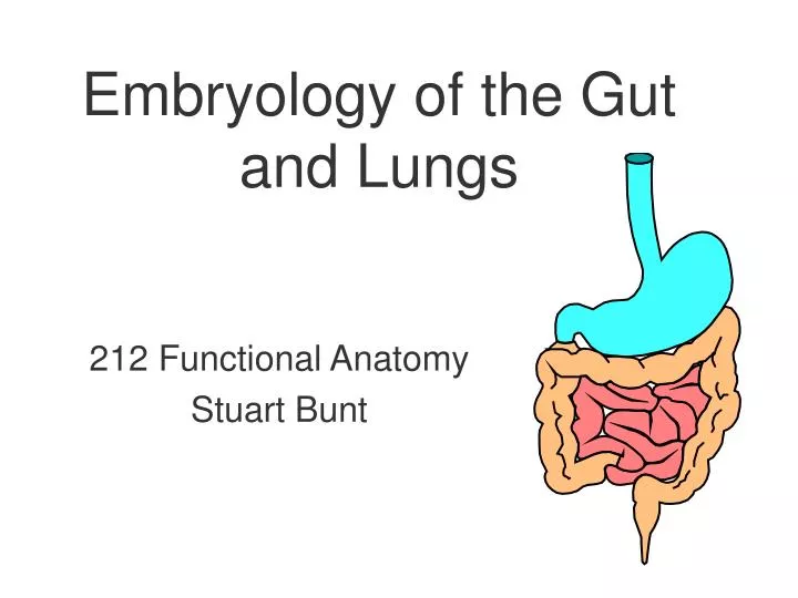
Embryology of the Gut and Lungs. 212 Functional Anatomy Stuart Bunt. Embryonic Curvature traps part of the yolk sac inside the embryo to form the gut. Embryology of Gut. Lung Buds from the Gut. Stages of Lung Development. Physiological hernia. The large liver takes up abdominal space

E N D
Embryology of the Gut and Lungs 212 Functional Anatomy Stuart Bunt
Embryonic Curvature traps part of the yolk sac inside the embryo to form the gut.
Physiological hernia • The large liver takes up abdominal space • Intestine lengthens into umbilicus • Rotates as it grows out and again as it re-enters abdomen • Omphalocele....no re-entry • Umbilical hernia, goes back out covered in peritoneum
Complex adult layout due to 270o rotation • Foregut, Midgut and Hindgut suspended by the dorsal mesentary, initially straight • Ventral mesentary connects stomach and ant. abd. wall, rest of gut free anteriorly • Mesentary supplies blood and nerves to gut between layers of peritoneum
Stomach rotates and distends Front Dorsal Mesentary Ventral Mesentary Back Splenic tissue Omentum Epiploic Foramen
Liver and spleen form in mesentary Anterior 1. Falciform ligament 2. Lesser Omentum 3. Dorsal mesentary, divided into:- 4a. Gastrosplenic ligament 4b. Lienorenal ligament 1 Liver 2 Stomach 4a Spleen 3 4b Posterior
Blood Supply of the Gut • Celiac trunk • Foregut • Midgut • Superior mesenteric artery • Hindgut • Inferior mesenteric artery • Rectum • Internal iliac artery (pudendal and rectal arteries)
Blood Supply to Abdominal Organs • Celiac trunk • Foregut • Midgut • Superior mesenteric artery • Hindgut • Inferior mesenteric artery • Rectum • Internal iliac artery (pudendal and rectal arteries)
Peritoneum • Flattened endothelial cells on fibro-elastic connective tissue • Parietal and visceral layers • Makes gut watertight • Suspends gut • Contains nerves and blood vessels • Omentum contains infection Flattened Endothelial cell
Mesenteries are important:- • Paracolic gutters channel fuid • Stop herniation due to bipedal posture • Supply blood/nerves • Sensitive to stretch • Contain infection • Useful in surgery
Intra and Retroperitoneal • To prevent the intestines falling into the pelvis our upright posture has been accompanied by a fusion of parts of the gut tube to the posterior abdominal wall. These parts become retro-peritoneal.
Uro-rectal septum divides the bladder and urogenital sinus from the rectum