Download
1 / 40
940 likes | 2.84k Views
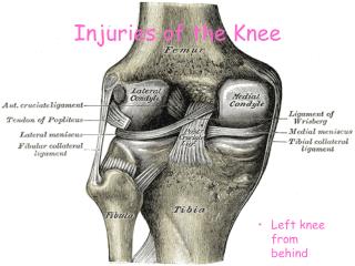
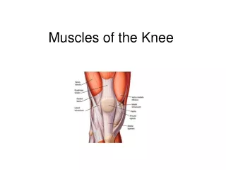
BASIC ULTRASOUND OF THE KNEE. By Mohamed Hassan Youssef MD Arthritis/ Rehab&Pain Clinic Board certified of ABPM&R. Knee Joint. Knee Bones. Distal end of the F umer Proximal end of the Tibia Head of the Fibula Patella. Knee Bones. Knee Bones. Movement of Patella Upward

E N D
BASIC ULTRASOUND OF THE KNEE By Mohamed Hassan Youssef MD Arthritis/Rehab&Pain Clinic Board certified of ABPM&R
Knee Bones • Distal end of the Fumer • Proximal end of the Tibia • Head of the Fibula • Patella
Knee Bones Movement of Patella Upward during Knee Flexion
Common Knee Ligaments • Ant Cruciate Ligament • Post Cruciate Ligament • Medial Collateral Ligament • Lateral Collateral Ligament • Arcuate Ligament • Coronary Ligament
Knee Menisci • Medial Meniscus • Lateral Meniscus
Knee Bursae Suprapatellar Prepatellar Infrapatellar PesAnserinus
Knee Bursae Semimembraneusus Bursa
US probes commonly used in MSK • Linear • Curved Linear
Relation of the needle to the US Probe • Long Axis Needle can be seen as a metal line approaching The target. • Short Axis Needle appear as a metal bright point in the target.
Positioning of the Knee duringUS guided needle injection • 1-put a roll under the knee to keep it in 25°- 35° of flexion. • 2-Use a linear Probe.
US of Knee • Move the linear probe in midline of the thigh thigh 10 cm above upper edge of patella. • Move the probe downward till the lower edge of the probe touch the Upper edge of the patella to elaborate the Quadriceps Tendon
References • http://www.ultrasoundpaedia.com/normal-knee/ • Tom Clark Ultrasound course • Ultrasound guided Musculoskeletal Procedures