Download
1 / 63
650 likes | 718 Views
Learn about the components, functions, and regulation of the digestive system. Explore anatomy from mouth to stomach, peristalsis, hormonal control, and common disorders. Enhance your knowledge of digestion with detailed insights.

E N D
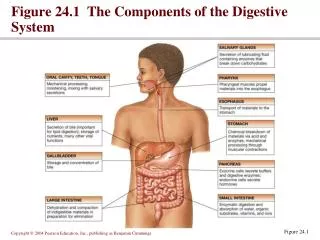
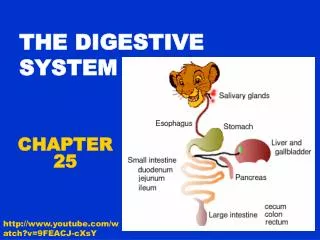
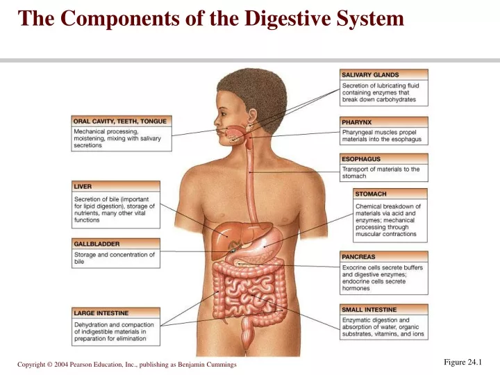
The Components of the Digestive System Figure 24.1
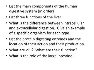
Functions of the digestive system • Ingestion • Mechanical processing • Digestion • Secretion • Absorption • Excretion
Movement of digestive materials • Visceral smooth muscle shows rhythmic cycles of activity • Pacemaker cells • Peristalsis • Waves that move a bolus • Segmentation • Churn and fragment a bolus
Peristalsis Figure 24.4
Control of the digestive system • Movement of materials along the digestive tract is controlled by: • Neural mechanisms • Hormonal mechanisms • Enhance or inhibit smooth muscle contraction • Local mechanisms • Coordinate response to changes in pH or chemical stimuli
]The Regulation of Digestive Activities Figure 24.5
The mouth opens into the oral or buccal cavity • Its functions include: • Analysis of material before swallowing • Mechanical processing by the teeth, tongue, and palatal surfaces • Lubrication • Limited digestion
The tongue • primary functions include: • Mechanical processing • Assistance in chewing and swallowing • Sensory analysis by touch, temperature, and taste receptors
The pharynx • Common passageway for food, liquids, and air • Lined with stratified squamous epithelium • Pharyngeal muscles assist in swallowing • Pharyngeal constrictor muscles • Palatal muscles
The Esophagus Figure 24.10a-c
Sequence Voluntary stage Push food to back of mouth Pharyngeal stage Raise Larynx + hyoid Tongue to soft palate Esophageal stage Contract pharyngeal muscles Open esophagus Start peristalsis Deglutition (swallowing)
Control Nerves Glossopharyngeal Vagus Accessory Brain stem Deglutition center Medulla oblongata Pons Disorders Dysphagia Aphagia Deglutition (swallowing)
Esophagus • Sphincters • Upper • Lower
Abnormalities • Achalasia – lower esophagus muscles don’t relax- doesn’t allow food to pass through • Atresia- esophogus openings are closed shut (Birth Defect) • Hernia- Esophogus pushes into stomach • Barret’s esophagus- tissue is more like intestinal lining tissue- can develop a rare cancer from this • Esophageal varices- enlarged veins that can leak blood
Functions of the stomach • Bulk storage of undigested food • Mechanical breakdown of food • Disruption of chemical bonds via acids and enzymes
Digestion and absorption in the stomach • Preliminary digestion of proteins • Pepsin • Permits digestion of carbohydrates • Very little absorption of nutrients • Some drugs, however, are absorbed • Mucous secretion containing several hormones • Enteroendocrine cells (Endocrine= Hormones!) • G cells secrete gastrin – stimulates pareital cells to secrete HCL • D cells secrete somatostatin- inhibits gastric secretions
The Stomach Figure 24.12b
The Stomach Lining Figure 24.13a, b
The Stomach Lining Figure 24.13c, d
Stomach Histology • Gastric pits: openings for gastric glands. Lined with simple columnar epithelium • Cells of gastric pits • Surface mucus: mucus that protects stomach lining from acid and digestive enzymes • Mucous neck: mucus • Parietal: hydrochloric acid and intrinsic factor • Chief: pepsinogen • Endocrine: regulatory hormones • Gastrin-containing cells: secrete gastrin (a hormone that stimulates acid secretion) • Somatostatin-containing cells: secrete somatostatin that inhibits gastrin and insulin secretion
Secretions of the Stomach • Chyme: ingested food plus stomach secretions • Mucus: surface and neck mucous cells • Viscous and alkaline • Protects from acidic chyme and enzyme pepsin • Irritation of stomach mucosa causes greater mucus • Intrinsic factor: parietal cells. Binds with vitamin B12 and helps it to be absorbed in the ileum. B12 necessary for DNA synthesis and RBC production (lack of B12 absorption leads to pernicious anemia)
Secretions of the Stomach • HCl: parietal cells • Kills bacteria (found in ingested food) • Stops carbohydrate digestion by inactivating salivary amylase • Denatures proteins • Helps convert pepsinogen to pepsin (optimal activity at pH 3 or less) • Pepsinogen: packaged and released by exocytosis. Pepsin catalyzes breaking of covalent bonds in proteins (breaks them into smaller peptide chains)
Cephalic Phase • The taste or smell of food, tactile sensations of food in the mouth, or even thoughts of food stimulate the medulla oblongata. • Parasympathetic action potentials are carried by the vagus nerves to the stomach, where enteric plexusneurons are activated. • Neurons stimulate secretion by parietal and chief cells (HCl and pepsin) and stimulate the secretion of the hormone gastrin and histamine. • Gastrin is carried through the circulation back to the stomach where it and histamine stimulate further secretion of HCl and pepsin.
GastricPhase • Distention of the stomach activates a parasympathetic reflex. Action potentials are carried by the vagus nerves tothe medulla oblongata. • Medulla oblongata stimulates further secretions of the stomach. • Distention also stimulates local reflexes that amplify stomach secretions.
IntestinalPhase • Chyme in the duodenum with a pH less than 2 or containing lipids inhibits gastric secretions by three mechanisms • Sensory input to the medulla from the duodenum inhibits the motor input from the medulla to the stomach. Stops secretion of pepsin and HCl. • Local reflexes inhibit gastric secretion • Secretin, and cholecystokininproduced by the duodenum decrease gastric secretions in the stomach.
Movements in Stomach • Combination of mixing waves (80%) and peristaltic waves (20%) • Both esophageal and pyloric sphincters are closed.
Small intestine • Important digestive and absorptive functions • Secretions and buffers provided by pancreas, liver, gall bladder • Three subdivisions: • Duodenum • Jejunum • Ileum • Ileocecal sphincter • Transition between small and large intestine
Figure 24.16 Regions of the Small Intestine Figure 24.16a
Histology of the small intestine • Plicae • Transverse folds of the intestinal lining • Villi • Fingerlike projections of the mucosa • Lacteals • Terminal lymphatic in villus • Intestinal glands • Lined by enteroendocrine, goblet and stem cells
Modifications to Increase Surface Area • Increase surface area 600 fold • Plicae circulares (circular folds) • Villi that contain capillaries and lacteals. Folds of the mucosa • Microvilli: folds of cell membranes of absorptive cells
The Intestinal Wall Figure 24.17a
The Intestinal Wall Figure 24.17b, c
The Intestinal Wall Figure 24.17d, e
Intestinal juices • Moisten chyme • Help buffer acids • Maintain digestive material in solution
Mucosa and Submucosa of the Duodenum • Cells and glands of the mucosa • Absorptive cells: cells with microvilli, produce digestive enzymes and absorb digested food • Goblet cells: produce protective mucus • Endocrine cells: produce regulatory hormones (Secretin, and cholecystokinin) • Granular cells (paneth cells): may help protect from bacteria (contain lysozymes) • Intestinal glands (crypts of Lieberkühn): tubular glands in mucosa at bases of villi [secrete sucrase ,maltase, trypsin, chymotrypsin, and pepsin (endopeptidases and exopeptidases) ] • Duodenal glands (Brunner’s glands): tubular mucous glands of the submucosa. Open into intestinal glands [produce a mucus-rich alkaline secretion (containing bicarbonate)
Jejunum and Ileum • Gradual decrease in diameter, thickness of intestinal wall, number of circular fold, and number of villi the farther away from the stomach • Major site of nutrient absorption • Peyer’s patches: lymphatic nodules numerous in mucosa and submucosa • Ileocecal junction: where ileum meets large intestine. Ileocecal sphincter (ring of smooth muscle) and ileocecal valve (one-way valve)
The Pancreas Figure 24.18a-c
Pancreatic Secretions: Pancreatic Juice • Enzymatic portion: (without the enzymes produced by pancreas, lipids, proteins, & carbs not adequately digested) • Trypsinogen- active form is trypsin--------proteolytic enzyme • Chymotrypsinogen- active form is chymotrypsin--------proteolytic enzyme • Procarboxypeptidase- active form is carboxypeptidase-------proteolytic enzyme • Pancreatic amylase- continues digestion of starch. • Pancreatic lipases- lipid digesting enzyme • Deoxyribonucleases and ribonucleases- reduce DNA & RNA to their nucleotide • Interaction of duodenal and pancreatic enzymes • Enterokinase is a proteolytic enzyme from the duodenal mucosa and it activates trypsinogen to trypsin. • Trypsin activates chymotrypsinogen to chymotrypsin. • Trypsin activates procarboxypeptidase to carboxypeptidase.
Liver Histology • Hepatic cords: radiate out from central vein. Composed of hepatocytes • Hepatic sinusoids: between cords, lined with endothelial cells and hepatic phagocytic (Kupffer) cells • Bile canaliculus: between cells within cords • Hepatocyte functions • Bile production • Storage • Interconversion of nutrients • Detoxification • Synthesis of blood components
Functions of the Liver • Bile production: 600-1000 mL/day. Bile salts, bilirubin (bile pigment that results from breakdown of hemoglobin), cholesterol, fats, fat-soluble hormones, lecithin • Neutralizes and dilutes stomach acid (neutralizes chyme so that pancreatic enzymes can function) • Bile salts emulsify fats. Most are reabsorbed in the ileum. (90% bile salts reabsorbed in the ileum & carried back to liver) • Secretin (from the duodenum) stimulates bile secretions, increasing water and bicarbonate ion content of the bile • Storage • Glycogen, fat, vitamins (A, B12, D, E, and K), copper and iron. Hepatic portal blood comes to liver from small intestine (nutrients are stored and secreted back into circulation when needed) • Synthesis • Blood proteins: Albumins, fibrinogen, globulins, heparin, clotting factors (liver produces its own new compounds)
Liver • Lobes • Major: Left and right • Minor: Caudate and quadrate • Porta: on inferior surface. Vessels, ducts, nerves, exit/enter liver • Hepatic portal vein, hepatic artery, hepatic nerve plexus enter • Lymphatic vessels, two hepatic ducts exit • Ducts • Right and lefthepatics (which transport bile out of liver) unite to form • Common hepatic • Cystic: from gallbladder • Common bile: union of cystic duct and common hepatic duct
The Anatomy of the Liver Figure 24.19b, c
Liver Histology Figure 24.20a, b
The gallbladder • Hollow, pear-shaped organ • Stores, modifies and concentrates bile
The Gallbladder Figure 24.21a, b
Large Intestine • Extends from ileocecal junction to anus • Consists of cecum, colon, rectum, anal canal • Movements sluggish (18-24 hours); chyme converted to feces. • Absorption of water and salts, secretion of mucus, extensive action of microorganisms are involved in the formation of feces. • 1500 mL chyme enter the cecum, 90% of volume reabsorbed yielding 80-150 mL of feces