Download
1 / 62
620 likes | 690 Views
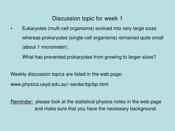
Discussion topic for week 1 Eukaryotes (multi-cell organisms) evolved into very large sizes whereas prokaryotes (single-cell organisms) remained quite small (about 1 micrometer). What has prevented prokaryotes from growing to larger sizes? Weekly discussion topics are listed in the web page:

E N D
Discussion topic for week 1 • Eukaryotes (multi-cell organisms) evolved into very large sizes whereas prokaryotes (single-cell organisms) remained quite small (about 1 micrometer). What has prevented prokaryotes from growing to larger sizes? Weekly discussion topics are listed in the web page: www.physics.usyd.edu.au/~serdar/bp/bp.html Reminder: please look at the statistical physics notes in the web page and make sure that you have the necessary background.
Basic properties of cells(Nelson, chap. 2) • Fundamental structural and functional units • Use solar or chemical energy for mechanical work or synthesis • Protein factories (ribosome) • Maintain concentration differences of ions, which generates a potential difference with outside (-60 mV) • Sensitive to temperature, pressure, volume changes • Respond to changes in environment via sensors and motility • Sense and respond to changes in internal conditions via feedback and control mechanisms (extreme example: apoptosis--cell death)
Two kinds of cells: • Prokaryotes (single cells, bacteria, e.g. Escherichia coli) Size: 1 mm (micrometer), thick cell wall, no nucleus The first life forms. Simpler molecular structures, hence easier to study Flagella: long appendages used for moving • Eukaryotes (everything else) Size: 10 mm, no cell wall (animals), has a nucleus, Organelle: subcompartments that carry out specific tasks e.g. mitochondria produces ATP from metabolism (the energy currency) chloroplast produces ATP from sunlight Cytoplasm: the rest of the cell
Molecular parts Electrolyte solution: water (70%) ions (Na+, K+, Cl-,…)
Organic molecules Hydrocarbon chains (hydrophobic) Double bonds
Functional groups in organic molecules Polar groups are hydrophilic. When attached to hydrocarbons, they modify their behaviour.
Four classes of macromolecules: polysaccharides, triglycerides, polypeptides, nucleic acids. (sugars) (lipids) (proteins) (DNA) Simple sugars (monosaccharides): e.g. ribose (C5H10O5),
Glucose is a product of photosynthesis Glucose and fructose have the same formula (C6H12O6) but different structure
Disaccharides are formed when two monosaccharides are chemically bonded together.
Lipids (fatty acids) are involved in long-term energy storage Saturated fatty acids Unsaturated fatty acid (C=C bonds)
Phospholipids are important structural components of cell membranes Phosphatide: At normal pH (7), the oxygens in the OH groups are deprotonated, leading to a negatively charged membrane. Phospatidylcholine (PC): The most common phospholipid has a choline group attached ….PO4-CH2-CH2-N+-(CH3)3
Proteins (polypeptides) perform control and regulatory functions (e.g. enzymes, hormones, ). The building blocks of proteins are the 20 amino acids.
Formation of polypeptides In water:
Protein structure 3.6 amino acids per turn, r=2.5 Å pitch (rise per turn) is 5.4 Å
Nucleic acids are formed from ribose+phosphate+base pairs The base pairs are A-T and C-G in DNA In RNA Thymine is substituted by Uracil
Adenosine triphosphate (ATP) has three phosphate groups. In the usual nucleotides, there is only one phosphate group which is called Adenosine monophosphate (AMP) Another important variant is Adenosine diphosphate (ADP)
B-DNA (B helix) ROM (Read-Only Memory) contains1.5 Gigabyte of genetic information Base pairs per turn (3.4 nm): 10
Primary structure of a single strand of DNA
Primary structure of a single strand of RNA
Hydrogen bonds among the base pairs A-T and C-G
Local structure of DNA Dynamic and flexible structure Bends, twists and knots Essential for packing 1 m long DNA in 1 mm long nucleus
Tools of Molecular Biology • X-ray diffraction • Nuclear magnetic resonance (NMR) spectroscopy • Electron microscopy • Atomic force microscopy • Mass spectrometry • Optical tweezers (single molecule exp’s) • Patch clamping (conductance of ion chanels) • Computational tools (molecular dynamics, bioinformatics, etc.) See, Methods in Molecular Biophysics by Serdyuk et al. for detailed discussion of these methods
Mass spectrometer Charged biomolecules are accelerated and injected to the velocity selector which has transverse E and B fields. Only those which have velocity v= E/B will pass through. In the next chamber, there is only a B field, which bends the beam by r = mv/Bq. The mass is accurately determined from the measured radius of gyration. Velocity selector
Optical tweezers Single molecule experiment using optical tweezers. Increasing the force on the bead triggers unfolding of RNA (Bustamante et al, 2001).
Patch clamping in ion channels (Neher & Sakmann) Using a clean pipette and suction, enable accurate measurement of picoamp currents in ion channels.
X-ray diffraction Basics 1. Accelerating charges emit radiation Larmor’s formula, non-relativistic • Where a is the acceleration of the charge and q is the angle between • the acceleration and radiation vectors. • Maximum radiation occurs in the direction perpendicular to a. • The only way to increase the intensity of radiation is via a. • Generic x-ray tubes use bremstrahlung (breaking radiation) • Isotropic, only selected wavelengths, low intensity • Synchrotrons accelerate electrons around a circular path (relativistic) • Directional, continuous, intense (one is operating in Melbourne now!)
2. Charged particles scatter incident radiation X-rays are electromagmetic radiation with l < 10 nm Where E is the the electric field amplitude, k is the wave vector and w is the frequency. An EM wave scattered by a charged particle has the amplitude Where q is the angle between incident and scattered radiation. Because nuclei are much heavier than electrons, they can be ignored. Note the q dependence; light atoms (e.g. H, He) are much harder to see.
Scattering from a collections of atoms is descibed using form factors Where r is the charge density, q is the momentum transfer in the scattering, i.e. q = k-k'. Thus form factor is just the Fourier transform of the charge density X-ray scattering provides information on f, which is then inverted via inverse Fourier transform to find the electron density maps
X-ray scattering from a single atom Atom in space 1D cut in FT 2D cut in FT
X-ray scattering from two atoms Braggs law: nl = 2d.sin(q) Atoms in space 1D cut in FT 2D cut in FT
X-ray scattering from 5 atoms in a row Atoms in space 1D cut in FT 2D cut in FT
X-ray scattering from a lattice of atoms Atoms in space reciprocal space
X-ray scattering from a monoclinic lattice (75 degrees) Atoms in space reciprocal space
X-ray scattering from a square box Atoms in space reciprocal space
X-ray scattering from a circular box Atoms in space reciprocal space