Download
1 / 46
460 likes | 657 Views
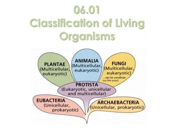
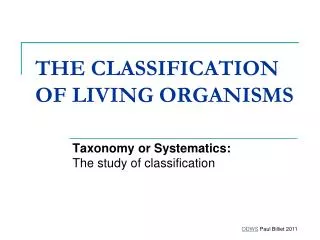
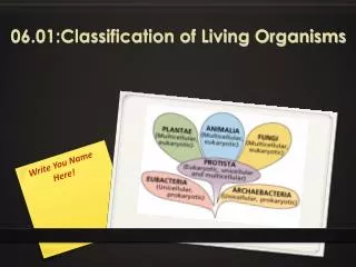
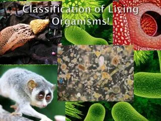
CLASSIFICATION OF LIVING ORGANISMS. Most Familiar organisms are made from Eukaryotic Cells e.g. plants animal fungi – including yeasts protists – protozoa (animal-like single celled) e.g. paramecia -algae (plant-like single/multi celled) e.g. algae.

E N D
Most Familiar organisms are made from Eukaryotic Cellse.g. plants animal fungi – including yeasts protists – protozoa (animal-like single celled) e.g. paramecia -algae (plant-like single/multi celled) e.g. algae
Plasma membrane Cytoplasm Mitochondrion Nuclear membrane Nuclear pore Chromatin Nucl;eolus Rough ER Ribosome Golgi Body Secretory vesicle nucleus Pinocytotic vesicle Centriole Food vacuole Smooth endoplasmic reticulum Lysosome) cytoplasm lysosome nucleolus vesicle
Vacuole membrane Large vacuole Smooth ER Plasmadesmata Secretory vesicle ribosome Chloroplast Cell membrane Mitochondrion Cell wall Nuclear membrane Nucleolus Chromatin Golgi body (dictyosome) Rough ER Nuclear pore
Plasmodesmata are channels found in plant cells which allow direct cytoplasmic connection between adjacent plant cells. • Middle Lamella –layer of “glue” between adjacent plant cells which holds them together. Contains pectins. • Nucleosome – DNA string wrapped around a histone protein bead. Necessary to allow DNA to be packaged efficiently into the small volume of the nucleus
Streptomyces coelicolor. The bacterium and its relatives produce most of the natural antibiotics in current use, including tetracycline and erythromycin. They also generate compounds that are used to treat cancer and suppress the immune system. Helicobacter pylori- Stomach uclers Mycobacterium tuberculosis – Tuberculosis Yersinia pestis – Black death (plague) Nesseiria meningitiis – Meningitis Bacteria 0.5-100m, Eukaryotes 10- 100 m
An Electron Micrograph of a bacterium Bacteria are much more simply constructed – no membrane bound organelles
COMMON BACTERIA SHAPES • Spherical (coccus) • Rod shaped (bacillus) • Spiral (spirochaetes, helicobacter)
cytoplasm pili (fimbriae) BASIC BACTERIAL STRUCTURES flagellae
Major difference between prokaryotic cells and eukaryotic cells No MEMBRANE BOUND ORGANELLES
PLASMA MEMBRANE Same as all membranes (bilipid layer) Phospholipid composition may differ between bacteria and eukaryotic cells.
The Cytoplasm • No compartmentalisation (no internal mebranes ie. No ER) • All chemical reactions occur within it. • Efficient regulation of biochemistry needed (Jacob Monod). • Contains ribosomes (free floating) responsible for protein synthesis – • different from eukaryotic ribosomes – • antibiotics (chloramphenicol, tetracycline, streptomycin) can specifically target them.
ON OFF OFF Repressor molecule No Enzyme is produced
ON ON ON Enzyme is produced LACTOSE
The Bacterial “Chromosome” - Nucleoid A single, circle of DNA. Packaged by folding – to reduce volume. FunctionsContains genetic information. Codes for bacterial proteins.
ReplicationBacteria reproduce by dividing (asexual) DNA must be replicated DNA simply copied (no MITOSIS - no chromosomes).
Extrachromosomal DNA – Plasmids StructureCircular DNA, smaller than nucleoid. Size~ 1000 - 200, 000 bp (c.f. 4,000,000 base pairs) 1-700 copies. FunctionNot normally essential, Gives some advantage e.g. antibiotic resistance. e.g. conjugative plasmids - Allow exchange of DNA between bacteria – antibiotic resistance can jump from one bacterial species to another.
The Cell Wall General Properties Cell wall resists swelling due to osmotic entry of water Prevents osmotic lysis Maintains shape Structure and synthesis unique to prokaryotes.
The chemical structure of peptidoglycan The NAM, NAG and amino acid side chain form PEPTIDOGLYCAN Covalently bonded (strong) to form a repeating polymer. The polymer is further strengthened by covalent cross links (peptide bridges) between amino acids. NAM – N acetyl muramic acid NAG – N acetyl glucosamine
Two basic types of bacterial cell wall structures – GRAM +ve • Gram +ve cells peptidoglycan is: • heavily cross-linked • very thick • (peptidoglycan accounting for 50% of weight of cell • and 90% of the weight of the cell wall) • 20-80 nm thick.
Two basic types of bacterial cell wall structures – GRAM -ve In GRAM –ve (G-) bacteria peptidoglycan much thinner 15-20% of the cell wall intermittently cross-linked.
GRAM STAINING Gram positive (G+) cells are purple and Gram negative (G-) cells are red.
Cell Wall Freely permeable to solutes, the openings in the mesh are large and all types of molecules can pass through them. Bleu D’avergne Lysozyme ( tears and saliva) -attacks peptidoglycan. It hydrolyzes the NAM - NAG linkage. Penicillin inhibits cells wall synthesis. The G+ cell wall is very sensitive to the action of lysozyme and penicillin. Penicillin is antibiotic of choice for infections caused by G+ organisms. e.g. Streptococcus pyrogenes which causes strep throat.
BACTERIAL MOTILITY Flagellum (ae) – used for movement
FIMBRIAE FLAGELLUM
Fimbriae/ pili –concerned with cell adhesion Special SEX pili – enable transfer of plasmids from one bacteria to another – can on occasion cross species. e.g staphylococcus - MRSA
SUMMARY No true membrane bound nucleus – rather a nucleoid (folded) No membrane bound organelles/ no compartmentalisation Many free floating ribosomes Cell walls made of peptidoglycan (G+/ G-) Mucilaginous capsule can be present Flagellae (movement) Pili./ Fimbrae are other extracellular protrusions (adhesion/ transfer)