Download
1 / 29
290 likes | 304 Views
Explore the four types of epithelial tissues (squamous, cuboidal, columnar, transitional) and their functions in histology. Learn about glandular epithelia and their classifications in this comprehensive guide to tissue study.

E N D
Tissue: The Living Fabric Part A 4
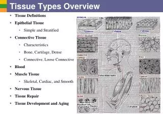
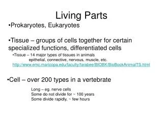
Tissues • Tissue is a group of cells similar in structure and function. • The study of tissues is called Histology. • The four types of tissues • Epithelial • Connective • Muscle • Nerve
Special Characteristics of the Epithelium: 1.) Cellularity – composed almost entirely of tight packed cells with very little extracellular space. 2.) Special contacts – form continuous sheets held together by tight junctions and desmosomes 3.)Polarity – apical and basal surfaces. 4.) Supported by connective tissue – reticular lamina and basal laminae Underneath the basal surface is a thin sheet of glycoproteins called the basal lamina. This sheet allows the cells to migrate toward a wound and is selectively permeable.
Special Characteristics of the Epithelium: • Avascular but innervated – contains no blood vessels but supplied by nerve fibers • Regenerative – rapidly replaces lost cells by cell
Classification of Epithelia • Simple or stratified Figure 4.1a
Classification of Epithelia • Squamous, cuboidal, or columnar Figure 4.1b
Epithelia: Simple Squamous • Single layer of flattened cells with disc-shaped nuclei and sparse cytoplasm • Functions • Diffusion and filtration • Provide a slick, friction-reducing lining in lymphatic and cardiovascular systems • Present in the kidney glomeruli, lining of heart, blood vessels, lymphatic vessels, and serosae
Epithelia: Simple Squamous Figure 4.2a
Epithelia: Simple Cuboidal • Single layer of cubelike cells with large, spherical central nuclei • Function in secretion and absorption • Present in kidney tubules, ducts and secretory portions of small glands, and ovary surface
Epithelia: Simple Cuboidal • Single layer of cubelike cells with large, spherical central nuclei • Function in secretion and absorption • Present in kidney tubules, ducts and secretory portions of small glands, and ovary surface Figure 4.2b
Epithelia: Simple Columnar • Single layer of tall cells with oval nuclei; many contain cilia • Goblet cells are often found in this layer • Function in absorption and secretion • Nonciliated type line digestive tract and gallbladder • Ciliated type line small bronchi, uterine tubes, and some regions of the uterus • Cilia help move substances through internal passageways
Epithelia: Simple Columnar Figure 4.2c
Epithelia: Pseudostratified Columnar • Single layer of cells with different heights; some do not reach the free surface • Nuclei are seen at different layers • Function in secretion and propulsion of mucus • Present in the male sperm-carrying ducts (nonciliated) and trachea (ciliated)
Epithelia: Pseudostratified Columnar • Single layer of cells with different heights; some do not reach the free surface • Nuclei are seen at different layers • Function in secretion and propulsion of mucus • Present in the male sperm-carrying ducts (nonciliated) and trachea (ciliated) Figure 4.2d
Epithelia: Stratified Squamous • Thick membrane composed of several layers of cells • Function in protection of underlying areas subjected to abrasion • Forms the external part of the skin’s epidermis (keratinized cells), and linings of the esophagus, mouth, and vagina (nonkeratinized cells)
Epithelia: Stratified Squamous • Thick membrane composed of several layers of cells • Function in protection of underlying areas subjected to abrasion • Forms the external part of the skin’s epidermis (keratinized cells), and linings of the esophagus, mouth, and vagina (nonkeratinized cells) Figure 4.2e
Epithelia: Stratified Cuboidal and Columnar • Stratified cuboidal • Quite rare in the body • Found in some sweat and mammary glands • Typically two cell layers thick • Stratified columnar • Limited distribution in the body • Found in the pharynx, male urethra, and lining some glandular ducts • Also occurs at transition areas between two other types of epithelia
Epithelia: Transitional • Several cell layers, basal cells are cuboidal, surface cells are dome shaped • Stretches to permit the distension of the urinary bladder • Lines the urinary bladder, ureters, and part of the urethra
Epithelia: Transitional • Several cell layers, basal cells are cuboidal, surface cells are dome shaped • Stretches to permit the distension of the urinary bladder • Lines the urinary bladder, ureters, and part of the urethra Figure 4.2f
Epithelia: Glandular • A gland is one or more cells that makes and secretes an aqueous fluid • Classified by: • Site of product release – • endocrine (internally secreting) • Exocrine (externally secreting) • Relative number of cells forming the gland • unicellular • multicellular
Endocrine Glands • Ductless glands that produce hormones. • Release of hormones is via exocytosis • Secretions include amino acids, proteins, glycoproteins, and steroids. • Endocrine glands vary in shape, size and structure.
Exocrine Glands • More numerous than endocrine glands • Secrete their products onto body surfaces (skin) or into body cavities • Examples include mucous, sweat, oil, and salivary glands
Unicellular Exocrine Gland • The only important unicellular gland is the goblet cell. • These single celled glands line our digestive, respiratory and intestinal tract. • They sit in between the columnar cells. • They produce mucin which breaks down in water to form mucus.
Multicellular Exocrine glands • Multicellular exocrine glands are composed of a duct and secretory unit. • Supportive Connective Tissue surrounds the gland and supplies it with blood vessels and nerve fibers.
Multicellular Exocrine Glands • Classified according to structure: • Simple or compound duct type • Tubular, alvelar or tubuloaveolar
Structural Classification of Multicellular Exocrine Glands Figure 4.3a-d
Structural Classification of Multicellular Exocrine Glands Figure 4.3e-g
Modes of Secretion • Multicellular glands secrete the chemical in different ways. • Exocytosis: this type of gland is called a merocrine gland. • Rupture: this type of gland has the cells fill with the chemical and then the whole cell ruptures. This type of gland is called holocrine gland.