Download
1 / 36
360 likes | 425 Views
Explore the history of DNA discovery, from Frederick Griffith's transformational bacteria research in 1928 to Watson and Crick's groundbreaking double helix model in 1953. Learn about Chargaff's rule, X-ray diffraction, base pairing, and the significance of DNA's structure in storing genetic information. Discover the secrets hidden within the DNA double helix and its fundamental role in genetics and heredity.

E N D
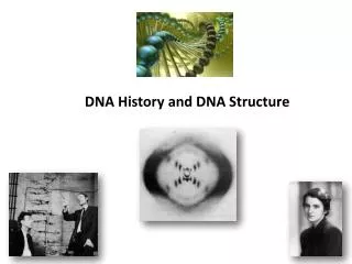
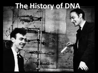
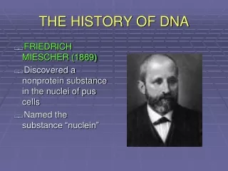
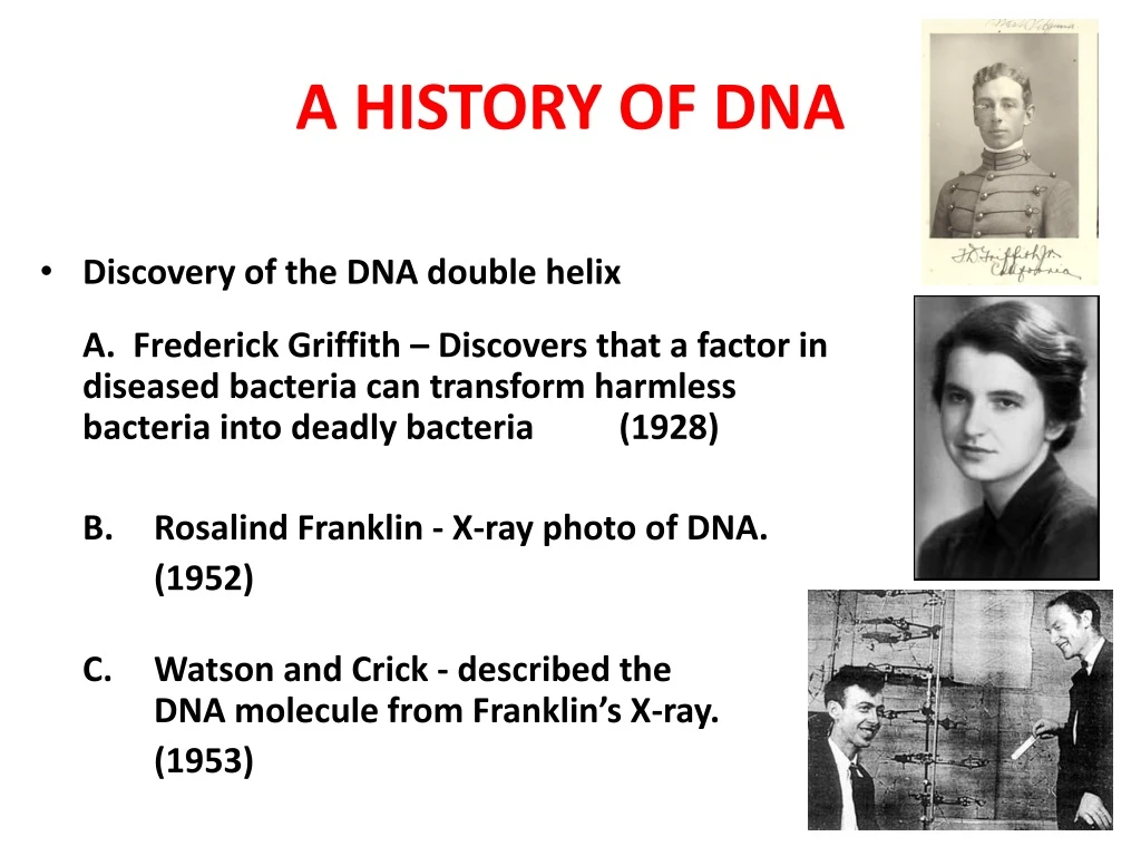
A HISTORY OF DNA • Discovery of the DNA double helix A. Frederick Griffith – Discovers that a factor in diseased bacteria can transform harmless bacteria into deadly bacteria (1928) B. Rosalind Franklin - X-ray photo of DNA. (1952) C. Watson and Crick - described the DNA molecule from Franklin’s X-ray. (1953)
Watson & Crick proposed • DNA had specific pairing between the nitrogen bases: • ADENINE – THYMINE • CYTOSINE - GUANINE • DNA was made of 2 long stands of nucleotides arranged in a specific way called the “Complementary Rule”
The Watson and Crick DNA Double helix • The correct structure of DNA was first deduced by J.D. Watson and F.H.C.Crick in 1953. • Their double helix model of DNA structure was based on two major kind of evidence. 1. Chargaff’s rule 2. X – ray diffraction patterns.
Chargaff’s rule • The composition of DNA from many different organisms was analyzed by E.Chargaff and his colleagues. • It was observed that concentration of thymine was always equal to the concentration of adenine (A = T) • And the concentration of cytosine was equal to the concentration of guanine (G = C). • This strongly suggest that thymine and adenine as well as cytosine and guanine were present in DNA with fixed interrelationship. • Also the total concentration of purines (A +G) always equal to the total concentration of pyrimidine (T +C). However, the (T+ A)/ (G+C) ratio was found to vary widely in DNAs of different species.
Adenine Thymine Guanine Cytosine The bases always pair up in the same way Adenine forms a bond with Thymine and Cytosine bonds with Guanine
X ray diffraction • When X rays are focused through isolated macromolecules or crystals of purified molecules, the X ray are deflected by the atom of the molecules in specific patterns called diffraction patterns. • It provides the information about the organization of the components of the molecules. • Watson and Crick had X ray crystallographic data on DNA structure from the studies of Wilkins and Franklin and their coworkers. • These data indicated that DNA was a highly ordered, multiple stranded structure with repeating sub structures spaced every 3.4 Ao (1 Angstrom = 10-10 m )
X-ray diffraction patterns of DNA – Rosalind Franklin and Maurice Wilkins The central cross shaped pattern as indicative of a helical structure. The heavy dark patterns (top and bottom) indicate that the bases are stacked perpendicular to the axis of the molecule.
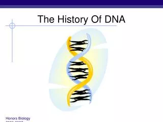
Double Helix • Watson and Crick proposed that DNA exists as a double helix in which two polynucleotide chains are coiled above one another in a spiral. • Each polynucleotide chain consists of a sequence of nucleotide linked together by Phosphodiester bonds. • The two polynucleotide strands are held together in their helical configurations by hydrogen bonding. • The base pairing is specific • That is, adenine is always paired with thymine and guanine is always paired with cytosine • Thus, all base-pairs consists of one purine and one pyrimidine. • Once the sequence of bases in one strand of DNA double helix is known, it is possible to know the other strand sequence of base because of specific base pairing.
In their most structural configuration, adenine and thymine form two hydrogen bonds, where as guanine and cytosine form three hydrogen bonds. • The two strands of a DNA are complementary (not identical) to each other. It is this property, that makes DNA uniquely suited to store and transmitting the genetic information. • The base-pairs in DNA are stacked 34Ao apart with 10 base-pairs per turn (3600) of the double helix • The sugar – phosphate backbones of the two complementary strands are antiparallel, that is they have opposite chemical polority.
As one move unidirectionally along a DNA double helix, the phosophodiester bonds in one bonds in one strand go from a 3’Carbon of one nucleotide to a 5’Carbon of the adjacent nucleotide. • Where as those in complementary strand go from 5’Carbon to a 3’carbon. • This opposite polarity of the complementary strands is very important in considering the mechanism of replication of DNA. • The high degree of stability of DNA double helices results in part from the large number of hydrogen bonds between base pairs.
Although each hydrogen bond by itself quite weak, since no. of hydrogen bonds are more, it can withstand. • The planar sides of the base pair are relatively non polar and thus tend to be water insoluble (hydrophobic). • The hydrophobic core stacked base-pairs contributes considerable stability to DNA molecules present in the aqueous protoplasms of living cells.
DNA • The amino acid sequence of a polypeptide is programmed by a gene. • A gene is a small region in the DNA. • Nucleic acids store and transmit hereditary information المعلومات الوراثية. • There are two types of nucleic acids: ribonucleic acid (RNA) and deoxyribonucleic acid (DNA). • DNA also directs mRNA synthesis, thus, controls protein synthesis. • Organisms inherit تتوارث DNA from their parents. • Each DNA molecule is very long and usually consists of hundreds to thousands of genes. • When a cell divides تنقسم, its DNA is copied and passed to the next generation of cells. • The mRNA interacts with ribosomes to direct the synthesis of amino acids in a polypeptide (protein)
The flow of genetic information is from DNA mRNA protein. • Protein synthesis occurs in ribosomes. • In eukaryotes, DNA is located in the nucleus, but most ribosomes are in the cytoplasm with mRNA as an intermediary وسيط.
NUCLEIC ACID STRUCTURE • DNA and RNA are large macromolecules with several levels of complexity • 1. Nucleotides form the repeating units • 2. Nucleotides are linked to form a strand • 3. Two strands can interact to form a double helix • 4. The double helix folds, bends and interacts with proteins resulting in 3-D structures in the form of chromosomes
Nucleotides • The nucleotide is the repeating structural unit of DNA and RNA • It has three components • A phosphate group • A pentose sugar • A nitrogenous base
These atoms are found within individual nucleotides • However, they are removed when nucleotides join together to make strands of DNA or RNA A, G, C or T A, G, C or U The structure of nucleotides found in (a) DNA and (b) RNA
Base + sugar nucleoside • Example • Adenine + ribose = Adenosine • Adenine + deoxyribose = Deoxyadenosine • Base + sugar + phosphate(s) nucleotide • Example • Adenosine monophosphate (AMP) • Adenosine diphosphate (ADP) • Adenosine triphosphate (ATP)
Base always attached here Phosphates are attached there
Nucleotides are covalently linked together by phosphodiester bonds • A phosphate connects the 5’ carbon of one nucleotide to the 3’ carbon of another • Therefore the strand has directionality • 5’ to 3’ • The phosphates and sugar molecules form the backbone of the nucleic acid strand • The bases project from the backbone
Bases 3 5 o o H H DNA nucleotide Adenine (A) CH2 CH2 Base Base 5 H H Purine H H H H H H 4 1 Guanine (G) 3 2 o o o o o o P P Cytosine (C) o o Pyrimidine Thymine (T) 5 3 Structures of nucleic acids (DNA & RNA) Phosphate group Deoxyribose 3 Sugar-phosphate backbone
The PO4 group of one nucleotide is attached to the sugar of the next nucleotide in line. • The result is a “backbone” of alternating phosphates and sugars, from which the bases starts.
5 3 Hydrogen bonds Cytosine (C) Guanine (G) Nitrogenous bases Thymine (T) Adenine (A) Purine Pyrimidines 5 3 Sugar-phosphate backbones Nitrogenous bases القواعد النيتروجينية Uracil (U)
DNA • Adenine (A) would form 2 hydrogenbonds only with thymine (T) • Guanine (G) would form 3 hydrogen bonds only with cytosine (C).
o H CH2 H Deoxyribose sugar (O on C2 is missed) H Ribose sugar (no missed O) H o H CH2 H H H H OH Single stranded nucleic acid Double stranded nucleic acid Bases: A, G, C, U Bases: A, G, C, T DNA & RNA Deoxiribo-Nucleic-Acid Ribo-Nucleic-Acid
The nucleic acid strand is a polymer of nucleotides • Nucleic acids are polymers of monomers called nucleotides. • Each nucleotide consists of three parts: a nitrogen base, a pentose sugar, and a phosphate group. • The nitrogen bases (rings of carbon and nitrogen) come in two types: Purines and Pyrimidines. • The pentose sugar joined to the nitrogen base is ribose in nucleotides of RNA and deoxyribose in DNA. • The only difference between the sugars is the lack نقص of an oxygen atom on carbon 2 in deoxyribose.
Polynucleotides are synthesized by connecting the sugars of one nucleotide to the phosphate of the next with a phosphodiester link. • This creates a repeating backbone of sugar-phosphate units with the nitrogen bases as appendages. • The sequence of nitrogen bases along a DNA or mRNA polymer is unique for each gene. • Genes are normally hundreds to thousands of nucleotides long. • The linear order of bases in a gene specifies يُحدد the order of amino acids (the monomers of a protein).
Inheritance is based on replication of the DNA double helix • An RNA molecule is single polynucleotide chain (single strand). • DNA molecules have two polynucleotide strands (double strand) that spiral around to form a double helix.
The sugar-phosphate backbones of the two polynucleotides are on the outside of the helix. • Pairs of nitrogenous bases (one from each strand) connect the polynucleotide chains with hydrogen bonds. • Most DNA molecules have thousands to millions of base pairs (bP).
Because of their shapes, only some bases are compatible متوافقة with each other. • Adenine (A) always pairs with thymine (T) and guanine (G) with cytosine (C). • With these base-pairing rules, if we know the sequence of bases on one strand, we know the sequence on the opposite المقابل strand. • The two strands are complementary مكملين لبعضهما. • During preparations for cell division each of the strands serves as a template قالب نسخ to order nucleotides into a new complementary strand. • This results in two identical copies نسختين طبق الأصل of the original double-stranded DNA molecule. • The copies are then distributed توزع to the daughter cells. • This mechanism ensures that the genetic information is transmitted to the new cells.
DNA backbone One nucleotide DNA Molecule A G C T A T C U U T C G A T A G Repeated Sugar - Phosphate Sugar–Phosphate-Base Polynucleotide DNA Double stranded RNA single stranded DNA mRNA