Download
1 / 54
540 likes | 730 Views
Gastrointestinal Part III. Jan Bazner-Chandler RN, MSN, CNS, CPNP. Intussusception. Telescoping of part of intestine into an adjacent distal portion. Bowden Text. Intussusception. Common cause of intestinal obstruction in children ages 3 to 25 months

E N D
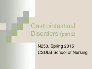
Gastrointestinal Part III Jan Bazner-Chandler RN, MSN, CNS, CPNP
Intussusception Telescoping of part of intestine into an adjacent distal portion. Bowden Text
Intussusception • Common cause of intestinal obstruction in children ages 3 to 25 months • Proximal segment of bowel prolapses or telescopes into the lumen of an immediately distal segment of bowel. • The prolapse results in vascular compromise, edema and mechanical obstruction. • Bleeding results in the passage of blood and mucous in the stool.
Assessment • Child appears with intermittent pain which is colicky, severe • Child will often draw legs up • Episodes occur 2-3 times / hour • Vomiting is prominent feature – bile stained vomiting a late sign • Bowel movements – bloody / mucous • Classic current jelly stool is a late sign
Clinical Manifestation Mucous + blood = classic “currant jelly” stool
Management • Reduce the obstruction before the bowel becomes necrotic. • Contrast Enema is diagnostic in 95% of cases and therapeutic curative in most cases. • Surgical reduction is radiologic reduction is not achieved.
Surgical Intervention • IV fluids + antibiotics pre-operatively. • Manual reduction is attempted. • If bowel perforation is noted during operative procedure a temporary colostomy may be needed.
Hirschsprung Disease • Congenital megacolon is a congenital absence of ganglio cells (ganglia). • This results in obstructed passage of stools causing the normal bowel to distend.
Assessment • In the newborn • No meconium in the first 24 hours • Bilious vomiting • Abdominal distention • Fever • Older infants / children • History of chronic constipation or fecal mass • Abdominal distention • Failure to thrive
Diagnosis and Treatment • Rectal biopsy • Removal of the aganglionic portion of the colon. • 1st stage surgery is often a colostomy • 2nd stage is pull-through surgery to connect the working colon to a point near the anus.
Colostomy at Birth Goal is to close colostomy and do “pull through” surgery at 6 to 12 months.
Long Term Complications • Anal stricture • Incontinence of stool • Short bowel syndrome
Pathophysiology • Inflammation of the vermiform appendix. • Obstruction at base blocks outflow of mucus. • Pressure builds up • Blood vessels are compressed. • Perforation and rupture
Assessment • Abdominal pain • Generalized to localized • Mc Burney’s point • Rebound tenderness • Loss of appetite • Vomiting • Low grade fever
Appendectomy Appendectomy
Ruptured Appendix Child develops high fever after a period of feeling better.
Perforation • Alert: With perforation of appendix, abdominal pain is suddenly relieved, but as peritonitis develops, it returns, along with signs of generalized acute abdomen. • Child will guard area of pain • Abdominal distension • High fever • May appear dehydrated
Interventions for Perforation • Extra fluids may be needed – a bolus of normal saline • NG may be inserted to decompress the stomach • IV antibiotics prior to surgical procedure • Fever control • Pain control
Post Operative Care Penrose drain acts as a wick to help drain the peroneal cavity after a ruptured appendix.
Interdisciplinary Interventions • Monitor I & O • Assess for bowel sounds • Dressing change as ordered • Ambulate ! Ambulate ! Ambulate ! • Cough and deep breath • Pain Management
GER or GERD • Effortless passage of gastric contents into the mouth (regurgitation) or esophagus. • Occurs in 1 in 500 infants • Higher incidence of GERD in esophageal atresia, neurologic impairment, cystic fibrosis, s/o omphalocele repair, chronic lung disease, bronchopulmonary dysplasia, asthma • By 1 year of age most infants with mild or moderate reflux are symptom free
GERD: Gastro-esophageal Reflux Disease • Infant older than 6 months, infant / child with congenital or neurological problems. • GER not relieved by simple measures. • Clinical Manifestations: • Regurgitation of feedings with slow growth / poor weight gain • Esophagitis = excessive crying • Apnea / Respiratory problems • Anemia
Diagnostic Tests • Barium swallow or Upper GI series • Gastric emptying study • Upper GI endoscopy • 24 hour pH monitoring
Conservative Management • Thickened formula • Frequent burping • Positioned with head elevated • after feeding • In infant with severe GERD positioning • prone with head of bed elevated
Surgical Management • Nissen fundoplication • Fundoplication creates a one • way valve by wrapping the • gastric fundus 360 degrees • around the lower end of • the esophagus • In severe cases may have • G-tube for 6-8 weeks after • surgery
Inflammatory Bowel Disease • A group of diseases characterized by inflammation of the GI tract. • Crohn’s Disease • Ulcerative Colitis
Inflammatory Bowel Disease • Refers to two chronic diseases that cause inflammation of the intestines. • Ulcerative Colitis • Crohn’s Disease
Causes • Most likely a genetic link that affects the immune system. • Incidence is increasing in both the pediatric and adult population • Prognosis depends on the type, severity and extent of the disease • Higher incidence of developing colorectal cancer
Ulcerative Colitis • Inflammatory disease of the large intestine. The inner lining or mucosa becomes inflamed, swells and ulcers develop. • Affects the lining of the bowel. • Most severe in the rectal area and anus.
Crohn’s Disease • Differs from ulcerative colitis in the areas of the bowel affected. • Most often affects the small intestine and parts of the large intestine. • Inflammation that extends deeper into the layers of the intestinal wall than ulcerative colitis.
Assessment • Ulcerative colitis: Bloody diarrhea with crampy, left-sided lower abdominal pain • Crohn’s disease: watery diarrhea with right lower quadrant pain • Both have • Weight loss • Anemia
Diagnostic Tests • Erythrocyte sedimentation rate ESR • Stool for gross or occult blood • Colonoscopy evaluation and biopsy • Genetic marker / family history
Pharmacologic Therapy • Corticosteroids during acute phase • Mesalazine – anti-inflammatory drug for mild to moderate cases. • Immunosuppression drugs: Azathioprine, methotrexate, 6-mercaptopurine • Remicade has been approved in severe cases
Long Term • Surgical removal of bowel if not managed by medical management. • Complications: • Alteration in body image due to steroids • Arthritis • Osteoporosis • Increase risk of colorectal cancer
Diagnostic Work-up for GERD • Upper GI series • Esophageal pH monitoring • Endoscopic exam
Medications to reduce symptoms including antacids or histamine-2 blocking agents Histamine 2 blocker: cimetadine Reglan or metaclopramide to enhance gastric emptying Pharmacologic Therapy
Necrotizing Enterocolitis Necrotizing = damage and death of cells Entero = refers to intestines Colitis = inflammation of the colon
NEC • 60 to 80% are premature infants • Feeding of concentrated formulas • Infants who have received blood transfusion • Infants with GI infections • Infants with polycythemia: congenital heart disease
Clinical Manifestations • History of formula feeding • Feedings stay in stomach • Abdominal distention / shiny abdomen • Bile-green fluid in stomach • Bloody bowel movements
Gas filled loops of bowel in NIC.
Interdisciplinary Interventions • NPO • Nasogastric tube to decompress stomach • IV fluid replacement • Antibiotics • Extra oxygen • Abdominal x-rays to monitor progress • Measure abdominal girth every four hours
Complications • Intestinal perforation • Surgery to remove dead bowel • Colostomy or ileostomy • Bowel is reconnected when infection and inflammation have resolved
Celiac Disease • Malabsorption caused by a life long intolerance to dietary gluten. • 1 in 100 children in USA • Genetic predisposition