Download
1 / 66
660 likes | 804 Views
The Skeleton Part A - Axial Skeleton. 7a. The Axial Skeleton. Eighty bones segregated into three regions Skull Vertebral column Bony thorax. The Skull. The skull, the body’s most complex bony structure, is formed by the cranium and facial bones

E N D
The Skeleton Part A - Axial Skeleton 7a
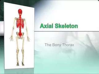
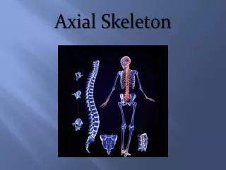
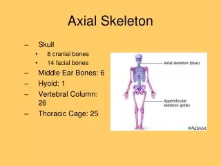
The Axial Skeleton Eighty bones segregated into three regions • Skull • Vertebral column • Bony thorax
The Skull • The skull, the body’s most complex bony structure, is formed by the cranium and facial bones • Cranium – protects the brain and is the site of attachment for head and neck muscles • Facial bones • Provide openings for the passage of air and food • Anchor the facial muscles of expression
Anatomy of the Cranium • Eight cranial bones – two parietal, two temporal, frontal, occipital, sphenoid, and ethmoid • Cranial bones are thin and remarkably strong for their weight
Anterior Aspects of the Skull Figure 7.2a
Parietal Bones and Major Associated Sutures Four sutures mark the articulations of the parietal bones • Coronal suture – articulation between parietal bones and frontal bone anteriorly • Sagittal suture – where right and left parietal bones meet superiorly • Lambdoid suture – where parietal bones meet the occipital bone posteriorly • Squamosal or squamous suture – where parietal and temporal bones meet
Inferior Portion of the Skull 9 8 7 6 5 3 2 4 1
Temporal Bones • Form the inferolateral aspects of the skull and parts of the cranial floor • Major markings include the zygomatic, styloid, and mastoid processes, and the mandibular fossae • Major openings include the stylomastoid and jugular foramina, the external and internal auditory meatuses, and the carotid canal
Temporal Bone Figure 7.5
Sphenoid Bone • Butterfly-shaped bone that spans the width of the middle cranial fossa • Forms the central wedge that articulates with all other cranial bones • Consists of a central body, greater wings, lesser wings, and pterygoid processes • Major markings: the sella turcica, hypophyseal fossa, and the pterygoid processes • Major openings include the foramina rotundum, ovale, and spinosum; the optic canals; and the superior orbital fissure
Sphenoid Bone Figure 7.6a, b
Ethmoid Bone • Most deep of the skull bones; lies between the sphenoid and nasal bones • Forms most of the bony area between the nasal cavity and the orbits • Major markings include the cribriform plate, crista galli, perpendicular plate, nasal conchae, and the ethmoid sinuses
Ethmoid Bone Figure 7.7
Facial Bones • Fourteen bones of which only the mandible and vomer are unpaired • The paired bones are the maxillae, zygomatics, nasals, lacrimals, palatines, and inferior conchae
Mandible and Its Markings • The mandible (lower jawbone) is the largest, strongest bone of the face • Its major markings include the coronoid process, mandibular condyle, and the mandibular and mental foramina
Mandible Figure 7.8a
Maxillary Bones • Medially fused bones that make up the upper jaw and the central portion of the facial skeleton • Facial keystone bones that articulate with all other facial bones except the mandible • Their major markings include palatine, frontal, and zygomatic processes, inferior orbital fissure, and the maxillary sinuses
Maxillary Bone Figure 7.8b
Zygomatic Bones • Zygomatic bones - Irregularly shaped bones (cheekbones) that form the prominences of the cheeks and the inferolateral margins of the orbits
Other Facial Bones • Nasal bones – thin medially fused bones that form the bridge of the nose • Lacrimal bones – contribute to the medial walls of the orbit • Palatine bones – two bone plates that form portions of the hard palate, the posterolateral walls of the nasal cavity
Other Facial Bones • Vomer – plow-shaped bone that forms part of the nasal septum • Inferior nasal conchae – paired, curved bones in the nasal cavity that form part of the lateral walls of the nasal cavity
Anterior Aspects of the Skull Figure 7.2a
Posterior Aspects of the Skull Figure 7.2b
External Lateral Aspects of the Skull Figure 7.3a
Midsagittal Lateral Aspects of the Skull Figure 7.3b
Inferior Portion of the Skull Figure 7.4a
Inferior Portion of the Skull 8 7 6 5 4 3 2 1 Figure 7.4b
Orbits • Bony cavities in which the eyes are firmly encased and cushioned by fatty tissue • Formed by parts of seven bones – frontal, sphenoid, zygomatic, maxilla, palatine, lacrimal, and ethmoid
Orbits Figure 7.9b
Nasal Cavity • Constructed of bone and hyaline cartilage • Roof – formed by the cribriform plate of the ethmoid • Lateral walls – formed by the superior and middle conchae of the ethmoid, the perpendicular plate of the palatine, and the inferior nasal conchae • Floor – formed by palatine process of the maxillae and palatine bone
Nasal Cavity Figure 7.10b
Paranasal Sinuses • Mucosa-lined, air-filled sacs found in five skull bones – the frontal, sphenoid, ethmoid, and paired maxillary bones • Lighten the skull and enhance the resonance of the voice
Paranasal Sinuses Figure 7.11
Hyoid Bone • Not actually part of the skull, but lies just inferior to the mandible in the anterior neck • Only bone of the body that does not articulate directly with another bone • Attachment point for neck muscles that raise and lower the larynx during swallowing and speech
Vertebral Column • Formed from 26 irregular bones (vertebrae) connected in such a way that a flexible curved structure results • Cervical vertebrae – 7 bones of the neck • Thoracic vertebrae – 12 bones of the torso • Lumbar vertebrae – 5 bones of the lower back • Sacrum – bone inferior to the lumbar vertebrae that articulates with the hip bones
Vertebral Column Figure 7.13
General Structure of Vertebrae • Body – disc-shaped, weight-bearing region • Vertebral arch – composed of pedicles and laminae that, along with the centrum, enclose the vertebral foramen • Vertebral foramina – make up the vertebral canal through which the spinal cord passes
General Structure of Vertebrae Figure 7.15
Cervical Vertebrae • Seven vertebrae (C1-C7) are the smallest, lightest vertebrae • C3-C7 are distinguished with an oval body, short spinous processes, and large, triangular vertebral foramina • Each transverse process contains a transverse foramen
CervicalVertebrae Table 7.2
The Atlas (C1) Figure 7.16a, b
The Axis (C2) • The axis has a body, spine, and vertebral arches as do other cervical vertebrae • Unique to the axis is the dens or odontoid process, which projects superiorly from the body and is cradled in the anterior arch of the atlas • The dens is a pivot for the rotation of the atlas
The Axis (C2) Figure 7.16c
The Atlas (C2) Figure 7.17a
Thoracic Vertebrae • There are twelve vertebrae (T1-T12) all of which articulate with ribs • Major markings include two facets and two demifacets on the heart-shaped body, the circular vertebral foramen, transverse processes, and a long spinous process • The location of the articulate facets prevents flexion and extension, but allows rotation of this area of the spine
Thoracic Vertebrae Figure 7.17b