Download
1 / 40
430 likes | 824 Views
Ms. Leonardo Roever. Multi-Slice CT for Coronary Calcium Scoring and Coronary Angiography. CT Angiogram Interpretation. Calcium Volume Score: ZERO CT angiography: Left Main, Circumflex, and Right coronary arteries: normal

E N D
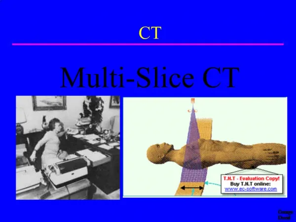
Ms. Leonardo Roever Multi-Slice CT for Coronary Calcium Scoring and Coronary Angiography
CT Angiogram Interpretation • Calcium Volume Score: ZERO • CT angiography: • Left Main, Circumflex, and Right coronary arteries: normal • LAD: eccentric, soft plaque adjacent to origin of first diagonal (~60% stenosis) • Correlation recommended
Cardiovascular Imaging - State of the Art • Multi-slice CT (MSCT) not likely to replace conventional angiography • Post-processing of images for MSCT angiography time & labor intensive • Major strength of CTA is its high negative predictive value • CMR to become the preferred cardiac imaging modality in the future
Which Test for Which Patient? • All modalities are improving • No single modality fits all applications and all patients • Choice of initial test depends on the specific clinical question in individual patient
4 to 64 Slice ScansFive Heart Beats 10 mm detector Pitch ~0.25 3 cm in 5 sec 20 mm detector Pitch ~0.25 6.2 cm in 5 sec 40 mm detector Pitch ~0.25 12.5 cm in 5 sec
Calcium Volume Scoring Area = 8 mm2 Peak CT = 290 Score = 8 x 2 = 16 Area = 15 mm2 Peak CT = 450 Score = 15 x 4 = 60 Total Score = S Hn x-factor (Agatston Scoring) 130-199 1 200-299 2 300-399 3 >400 4
The Calcium Scale The calcium scale is a linear scale with 4 calcium score categories: 0 none 1–99 mild 100–400 moderate >400 severe *Calcium score correlates directly with risk of events and likelihood of obstructive CAD*
Ethnic Differences in Coronary CalcificationThe Multi-Ethnic Study of Atherosclerosis (MESA) 6814 men and women aged 45-84 years Bild DE et al. Circulation. 2005;111:1313-1320.
Five-Year Mortality Rates in Framingham Risk Subsets by Coronary Calcium Score * *p<0.001 * * Shaw et al. Radiology 2003; 228:826-833
Progression of Coronary Artery Calcium and Risk of First MI495 Asymptomatic Patients Started on Statin Therapy • MI in 41 pts during 3.2 + 0.7 years • LDL levels similar in MI and non-MI pts • Relative risk of MI in presence of CAC progression was 17.2-fold higher (P<0.0001) Raggi P et al. Arterioscler Thromb Vasc Biol. 2004;24:1272-77.
Coronary Disease Progression Calcified Plaque Detected by CT >60% stenosis (+) stress/imaging Role for CTA ?
CTA Limitations • Rapid (>80 bpm) and irregular HR • High calcium scores (>800-1000) • Stents • Contrast requirements (Cr > 2.0 mg/dl) • Small vessels (<1.5 mm) and collaterals • Obese and uncooperative patients • RADIATION EXPOSURE
Clinical Indications for MSCT • Calcium Scoring (CS) - risk stratification in the intermediate risk patient • Non-invasive coronary angiography (CTA) in the symptomatic low-risk patient or asymptomatic intermediate-risk patient *A negative test (normal CTA) has a 98% chance of revealing normal coronary arteries on invasive angiography*
When to Consider MSCT • Equivocal stress test or persistent symptoms despite negative stress test • Prior to non-coronary cardiac surgery (valve or congenital repair) • Patients with difficult access or on therapeutic warfarin • Suspected coronary anomalies
CFX LAD RCA Lt Main
Pulmonary Vein Stenosis Vasamreddy et al. HeartRhythm (2004) 1, 78-81.
Aortic Coarctation Visualized by 16-Row Detector MSCT Fröhlich, G et al. Circulation. 2005;112:e81.
Pericardial CalcificationMulti-Slice CT Scanning Superior to MRI Hoffmann et al. Circulation 108 (7): 48e Figure IG1
Future Indications Nikolaou et al. Cardiology Clinics. 21;(2003):639-655.