Download
1 / 18
260 likes | 1.44k Views
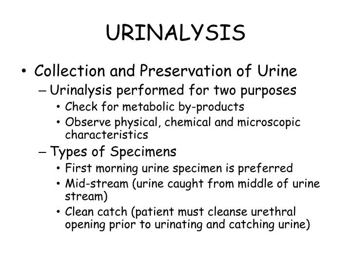
URINALYSIS. Collection and Preservation of Urine Urinalysis performed for two purposes Check for metabolic by-products Observe physical, chemical and microscopic characteristics Types of Specimens First morning urine specimen is preferred

E N D
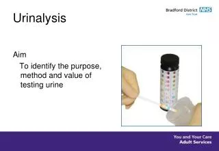
URINALYSIS • Collection and Preservation of Urine • Urinalysis performed for two purposes • Check for metabolic by-products • Observe physical, chemical and microscopic characteristics • Types of Specimens • First morning urine specimen is preferred • Mid-stream (urine caught from middle of urine stream) • Clean catch (patient must cleanse urethral opening prior to urinating and catching urine)
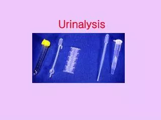
Handling and preserving specimens • Examine within 1 hour of collection OR • Refrigerate at 4-6° C for up to 8 hours • Preservatives (least ideal) • When urine sits at room temperature • Bacteria multiply • Glucose decreases • Casts and cellular elements decompose
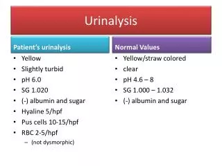
Color of urine • Yellow – dilute urine is usually lighter in color; concentrated urine is usually dark • Red – may have blood present • Brown/black – may be associated with melanoma • Yellow-brown or green-brown – may be associated with liver conditions such as hepatitis or cirrhosis
Transparency • Urine normally transparent • Turbid – may be associated with crystals that settle out of urine at room or refrigerator temperature • Cloudy – may be associated with UTI OR crystals
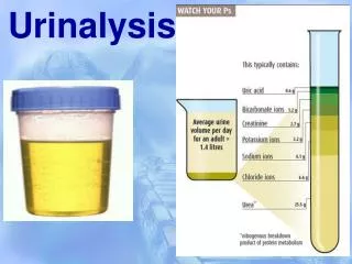
Unit #5A – Clinical Laboratory Testing - Urinalysis • Specific gravity is the ratio of the weight of a given volume of the solution (urine) to the weight of an equal volume of water • Indicates concentrations of dissolved chemicals such as glucose, salts, etc. • The result of the kidneys’ ability to concentrate urine • Normal values – 1.005-1.030 (Ave = 1.015) • Usually measured by dip stick or refractometer
Unit #5A – Clinical Laboratory Testing - Urinalysis • Chemical Examination of Urine • Reagent strips • Test pads are for pH, protein, glucose, ketone, bilirubin, blood, urobilinogen, specific gravity, leukocytes and bacteria • Used only once and discarded • Performing the chemical tests by reagent strip • Perform within 1 hour after collection OR • Allow refrigerated specimens to return to room temperature • Dip strip in fresh urine and compare color of pads to the color chart after appropriate time period • Instruments are available which detect color changes electronically
Unit #5A – Clinical Laboratory Testing - Urinalysis • Urine Multistix – reading dipstick results manually; colors are matched to those on the bottle label; timing is critical for each pad.
Bayer Clinitek automatically reads a urine dipstick and prints out results
Principle of chemical tests • pH measures degree of acidity or alkalinity of urine • Presence of protein (proteinuria) is an important indicator of renal disease, such as pyleonephritis • Presence of glucose (glycosuria) indicates that the blood glucose level has exceeded the renal threshold, such as in diabetes
Ketones are excreted when the body metabolizes fats incompletely (ketonuria), such as in diabetes • Bilirubin is a byproduct of the breakdown of hemoglobin. Its presence may be an indication of liver disease, bile duct obstruction or hepatitis • Presence of blood may indicate infection, trauma to the urinary tract or bleeding in the kidneys • Urobilinogen is a degradation product of bilirubin formed by intestinal bacteria. It may be increased in hepatic disease or hemolytic disease
Nitrite formed by gram negative bacteria converting urinary nitrate to nitrite. Presence of nitrites in fresh urine can indicate infection – in an old urine, nitrites can be positive without an infection • Leukocytes usually indicate infection • Specific gravity reflects kidney's ability to concentrate urine • Normal values • Negative results for glucose, ketones, bilirubin, nitrites, leukocyte esterase and blood • Protein negative or trace • pH 5.5-8.0
Microscopic Examination • Oraganized • RBC • WBC • Microorganisms • Yeast, Fungi and Bacteria • Epithelial Cells Yeast Fungi Bacteria RBC WBC Epithelial Cells
Microscopic Examination • Unorganized Sediment • Fat Globules • Precipitated Crystals • Acidic • Akaline Fat Globules in Urine
Acidic Crystals Tryosine Calcium Oxalate Dihydrate Cystine Amorphous Urates (Na, K, Mg, or Ca salts) Sodium Urate Leucine Uric Acid
Alkaline Crystals Amorphous Phosphate Ammonium Biurate Triple phosphates (Struvite) Dicalcium Phosphate Calcium Carbonate