Download
1 / 30
300 likes | 356 Views
This detailed text covers the anatomy of the larynx, including various cartilages, muscles, ligaments, and mucosal linings. Learn about the structures, functions, and clinical aspects of the larynx in a comprehensive manner.

E N D
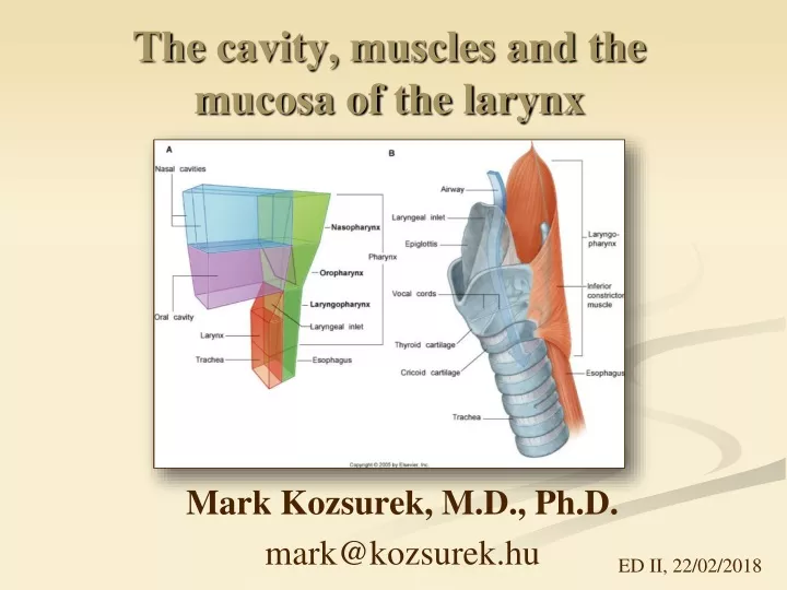
The cavity, muscles and the mucosa of the larynx Mark Kozsurek, M.D., Ph.D. mark@kozsurek.hu ED II, 22/02/2018
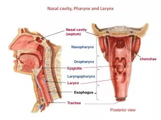
Triticeal cartilage Epiglottis Cuneiform cartilages [Wrisberg’s] Corniculate cartilages [Santorini’s] Arithenoid cartilage Thyroid cartilage Cricoid cartilage
Lat. thyrohyoid lig. Median thyrohyoid lig. Thyrohyoid membrane Lat. thyrohyoid lig. Hyoepiglottic lig. Thyroepiglottic lig. Vestibular lig. Vocal lig. Median cricothyroid lig. (conic lig.) Cricopharyngeal lig.
A now we put muscles onto this membranous-cartilagineus frame!
Thyroarythenoid* (Inter)- arythenoid Lat. crico-arythenoid Post. cricoarythenoid Cricothyroid Cricothyroid *: individual parts: Aryepiglottic, Thyroepiglottic, Oblique thyroarythenoid
Laryngeal muscles do not exclusively open or close the rima glottidis! – the most of the students only know these two possibilities! Vocal fold is stretched by the Cricothyroid and is relaxed by the Vocalis and Thyroarythenoid muscles. The Thyroarythenoid - especially during swallowing - also constricts the supraglottic part of the larynx, thus, acts as a sphincter muscle.
The only muscle which opens the rima glottidis is the Post. cricoarythenoid. Membranous part of the rima glottidis is closed by the Lat. cricoarythenoid, while the cartilagineous part requires the action of (Inter)arythenoid muscles for complete closure!
Lining the membranous-cartilagineus-muscular frame with mucosal membrane inside
Note: • Laminae of thethyroidcartilagefuseanteriorly, whilethelaminaofthecricoidcartilage is foundposteriorly! • The onlymuscle of thelarynxpassingoverthemidline is the (Inter)arythenoid, sothisistheonlyoneobservableinsagittalsection! quadrangular membrane: vestibular and aryepiglottic folds, lateral margin of epiglottis and the arythenoid cartilage (2 folds and two cartilages!) conus elasticus (triangular membrane taken twice): vocal fold, superior margin of cricoid cartilage, median cricothyroid lig.
Note: • Boundaries of thefibroelasticmembranes, • thecut (Inter)arythenoidmuscle, • sagittalycutlamina of cricioidcartilage, • and Vocalismuscletogetherwiththeobliqueandstraightparts of theCricothyroidmuscleaswellasthemediancricothyroid ligament aftertheremoval of themucosa and theconuselasticus!
frontal aspect vestibule ventricle subglottic space Identify cross sections of muscles seen on the image!
Thyroarythenoid, aryepiglottic part Thyrohyoid Vocalis Superior pharyngeal constrictor Lat. cricoarythenoid Sternothyroid Cricothyroid
ext. carotid artery → sup. thyroid artery → sup. laryngeal artery subclavian artery →thyrocervical trunk → inf. thyroid artery → inf. laryngeal artery
superiorly: identical to the arterious blood supply inferiorly: toward the internal jugular and left brachiocephalic veins
VAGUS NERVE sup. laryngeal nerve: int. branch: supraglottic mucosa ext. branch: Cricothyroid muscle inf. laryngeal nerve: (terminal br. of recurrent laryngeal n.) infraglottic mucosa and all the other muscles not innervated by the superior laryngeal nerve ACCESSORY NERVE mainly contributes to the innervation of the Interarythenoid muscles
ambiguusnucl.branchialmotor dorsalvagusnucl. generalvisceromotor spinaltrigeminalnucl. generalsomatosensory lateralnucl. of alacinereageneralviscerosensory solitarynucl. specialviscerosensory (taste)
Respiratory epithelium (pseudostratified columnar epithelium with kinocilia) and goblet cells. According to the increased mechanical demand vocal folds are lined by stratified squamous non-keratinized epithelium! Skeletal muscle, hyalin cartilage, elastic cartilage, mixed glands, vessels and nerves.
Conicotomy We arrive into the subglottic space after cutting the conic (median cricothyroid) ligament!