Download
1 / 46
460 likes | 672 Views
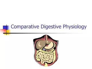
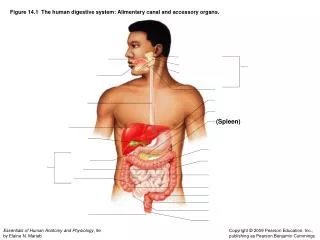
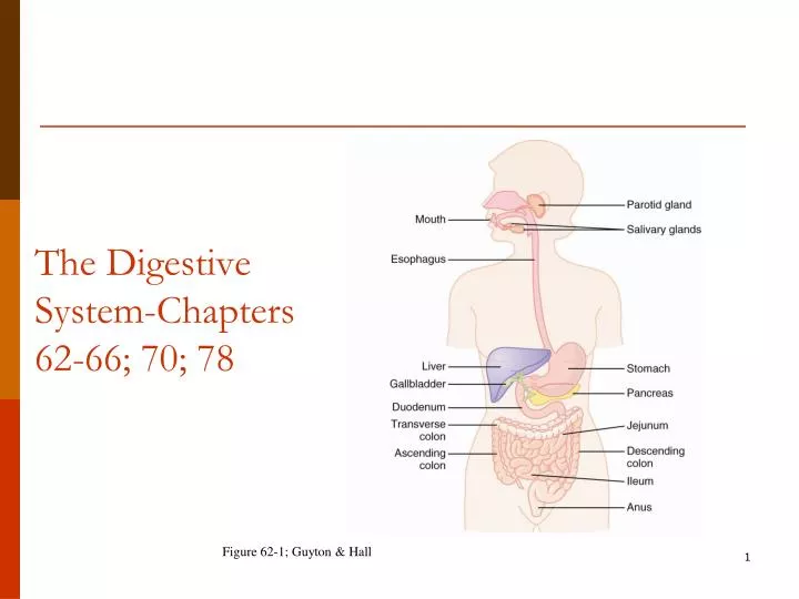
The Digestive System-Chapters 62-66; 70; 78. Figure 62-1; Guyton & Hall. Digestive Processes. Ingestion Propulsion Digestion: Mechanical and Chemical digestion Absorption- nutrients and water Defecation. Layers Alimentary Canal. 1. Serosa 2. Longitudinal muscle (muscularis externa)

E N D
The Digestive System-Chapters 62-66; 70; 78 Figure 62-1; Guyton & Hall
Digestive Processes • Ingestion • Propulsion • Digestion: Mechanical and Chemical digestion • Absorption- nutrients and water • Defecation
Layers Alimentary Canal • 1. Serosa • 2. Longitudinal muscle (muscularis externa) • 3. Myenteric (Auerbach’s) nerve plexus • 4. Circular muscle • 5. Submucosa • 6. Submucosal (Meissner’s) nerve plexus • 7. Muscularis mucosae • 8. Mucosa • 9. Epithelial lining
Autonomic nerve fibers • Both divisions found in myenteric and submucosal nerve plexi—What do they do? • Sensory neurons that monitor tension, and efferent visceral motor fibers. OWN SYSTEM! • Myenteric-GI motility control • Stimulatory influences - • tonic contraction (tone) • contraction frequency / intensity ( propulsion) • Inhibitory influences • Decreased Sphincter tone (relax) - pyloric sphincter, ileocecal sphincter, LES • Submucosal- Local control • Secretion • Absorption • Contraction of muscularis mucosa
Control of the digestive system • Movement of materials along the digestive tract is controlled by: • Neural mechanisms • Parasympathetic (Ach) and local reflexes • Hormonal mechanisms • Enhance or inhibit smooth muscle contraction • Local mechanisms • Coordinate response to changes in pH or chemical stimuli and stretching
Digestive Enzymes Intestinal Mucosa enterokinase sucrase maltase lactase amino-oligopeptidase dipeptidase Salivary glands -amylase lingual lipase Stomach pepsin Pancreas amylase trypsin chymotrypsin carboxypeptidase lipase cholesterolesterase
The mouth opens into the oral or buccal cavity Primary Secretion Alpha-amylase • Its functions include: • Analysis of material before swallowing • Mechanical processing by the teeth, tongue, and palatal surfaces • Lubrication • Limited digestion • Lingual lipase (negligible fat digestion) • Salivary amylase (limited carbohydrate digestion) • Antibodies and proteolytic enzymes
Digestion and absorption in the stomach • Short-term storage reservoir • Secretion of intrinsic factor • Pepsinogen • gastrin • Chemical and enzymatic digestion is initiated, particularly of proteins • Liquefaction of food • Slowly released into the small intestine for further processing
Gastric glands • Two types glands - • Gastric - HCl (oxyntic)pepsinogen intrinsic factor mucus • Pyloric - gastrin mucus
80% Gastric glands- 3 types of cells • Mucous Neck cell (goblet)- release mucus to protect mucosa from acid and pepsin • Parietal cells- HCl and intrinsic factor (B12 absorption by small intestine). • Chief- numerous and release pepsinogen
LUMEN BLOOD + H+ HO- H2O CO2 CO2 C.A. Final Results HCl - 155 mEq/L KCl - 15 mEq/L NaCl - 3 mEq.L pH = 0.8 P HCO3 HCO3 H+ K+ K+ K+ K+ H2O P Na+ P Na+ Na+ Na+ Cl- P Cl- Cl- Cl- H2O osmosis Acid production and secretion
20% 2 cell types of Pyloric gland • G-cells - release gastrin • Enteroendocrine cells -stimulates parietal cells to secrete acid and increases pyloric contraction; relaxes pyloric sphincter • Mucus neck cells - mucous
Weakens H. pylori, aspirin, ethanol, NSAIDs, bile salts Strengthens mucus, HCO3- secretion, gastrin, PGs, epidermal growth factor Gastric and Duodenal ulcers • Peptic ulcers occur when damaging effects of acid and pepsin overcome ability of mucosa to protect itself • Gastric ulcers - main problem is decreased ability of mucosa to protect itself • Duodenal ulcers - main problem is exposure to increased amounts of acid and pepsin
What is the Gastric Mucosal Barrier? • alkaline mucus resists the acid and enzymes • Tight junctions-gastric juice can’t seep into lamina propria • Epithelial cell replacement- 3-6 day life span. • Physiological - diffused H+ ions are transported back to lumen Damaged Gastric Mucosal Barrier • H+ back-leaks into mucosa in exchange for Na+. This is a forerunner to gastric ulcer - • Decreased cell pH leads to cell death • Damaged mast cells (ECL cells) leak histamine • Viscous cycle - Histamine .. vascular damage .. local ischemia .. greater leakage of H+.. more cell death ...
Helicobacter pylori • H. pylori found in 95% patients with DU and 100% patients with GU (when alcohol, aspirin, NSAIDS are eliminated) • Gram negative bacterium • High urease activity - high NH4+ activity - can withstand acid environment - NH4+ damages epithelial cells (GU) - Increases acid secretion (DU)
Treatment of Peptic Ulcers • Antacids • H2 receptor blockers - Rantidine (Zantac) - Cimetidine (Tagamet) • Proton pump inhibitors - Omeparazole (Prilosec) • Antibiotics • Surgical (rare) - vagotomy - antrectomy
Stimulation of acid secretion Seeing, smelling and anticipating food is perceived in brain. Brain tells stomach to prepare for receipt of meal Accounts for 30% of acid response to meal • Gastric secretion is stimulated by local (distention), neural, and endocrine mechanisms • Acetylcholine - HCl secretion • - mucus, pepsinogen, and gastrin • Histamine - HCl secretion • Gastrin - HCl secretion (1500x more powerful compared to histamine) 60% 10%
Small intestine • Important digestive and absorptive functions • Secretions and buffers provided by pancreas, liver, gall bladder • Three subdivisions: • Duodenum • Jejunum • Ileum • Ileocecal sphincter • Transition between small and large intestine
Plicae Transverse folds of the intestinal lining Villi Fingerlike projections of the mucosa Lacteals Terminal lymphatic in villus Microvilli Brush border: increases surface area 20-fold Histology of the small intestine
Intestinal glands • secretin to stimulate pancreas to release bicarbonate mucus • cholecystokinin to stimulate pancreas and gallbladder • Gastric Inhibitory peptide (GIP)- inhibits gastrin secretion and decreases stomach emptying • Duodenal glands- bicarbonate mucus.
The Activities of Major Digestive Tract Hormones Figure 24.22
Small Intestine- digestive enzymes • Maltase- splits maltose into 2 glucose units • Lactase- splits lactose into glucose and galactose • Sucrase- splits sucrose into glucose and fructose • Peptidase- breaks down small peptides into amino acids • Intestinal lipase- breaks down triglycerides into free fatty acids and monoglycerides • Enterokinase- Activates trypsinogen to trypsin (trypsin then activates chymotrypsinogen and procarboxypeptidase)
Pancreas • As chyme floods into small intestine two things must happen: • Acid must be neutralized to prevent damage to duodenal mucosa • Macromolecular nutrients - proteins, fats and starch must be broken down much further so their constituents can be absorbed • Pancreas plays vital role in accomplishing both objectives • Digestive enzymes for all food types • Bicarbonate solution to neutralize acid chyme
Regulation of Pancreatic Secretion • Secretin and CCK are released when fatty or acidic chyme enters the duodenum • CCK and secretin enter the bloodstream • Upon reaching the pancreas: • CCK induces the secretion of enzyme-rich pancreatic juice • Secretin causes secretion of bicarbonate-rich pancreatic juice • Vagal stimulation also causes release of pancreatic juice
The Pancreas • Exocrine function (98%) • Acinar cells make, store, and secrete pancreatic enzymes • Endocrine function – • ( cells) release somatostatin (inhibitory to gastrin and insulin and glucagon) • β-cells –release insulin • α-cells-Release glucagon
The Pancreas as an Endocrine Gland • Insulin • Beta cells • Skeletal muscle and adipose tissue need it to make glucose receptors • Promotes glucose uptake • Prevents fat and glycogen breakdown and inhibits gluconeogenesis • Increases protein synthesis • Promotes fat storage Epi/Norepi inhibit insulin! Help maintain glucose levels during times of stress and increase lipase activity in order to conserve glucose levels Picture from:http://www.dkimages.com/discover/Home/Health-and-Beauty/Human-Body/Endocrine-System/Pancreas/Pancreas-1.html
The Pancreas as an Endocrine Gland • Glucagon • Maintains blood glucose between meals and during periods of fasting. • Nervous tissue (brain) do not need insulin; but are heavily dependent on glucose levels! • Increases blood glucose levels. • Initiates glycogenolysis in liver (within minutes) • Stimulates amino acid transport to liver to stimulate gluconeogenesis Image from: http://www.dkimages.com/discover/previews/768/74261.JPG
Disorders of the Pancreas: Diabetes Mellitus • Gestational Diabetes • Type I diabetes – develops suddenly, usually before age 15 • Destruction of the beta cells • Skeletal tissue and adipose cells must use alternative fuel and this leads to ketoacidosis • Hyperglycemia results in diabetic coma
Disorders of the Pancreas: Diabetes Mellitus • Type II diabetes and metabolic syndrome– adult onset • Usually occurs after age 40 • Cells have lowered sensitivity to insulin • Controlled by dietary changes and regular exercise
Pancreatic Failure • Digestion is abnormal when pancreas fails to secrete normal amounts of enzymes. • Pancreatitis • Removal of pancreatic head - malignancy • Without pancreatic enzymes - • 60% fat not absorbed (steatorrhea) • 30-40% protein and carbohydrates not absorbed
Pancreatitis • Pancreatitis means inflammation of pancreas. Autodigestion theory can explain condition. • Chronic pancreatitis - (multiple shared causes) • alcohol - most common cause in adults • cystic fibrosis - most common cause in childre • CF patients lack chloride transporter at apical membrane. • Watery ductal secretion decreases which concentrates acinar secretions in ducts. • Precipitation of proteinaceous secretions block ducts and can destroy gland by autodigestion. • Acute pancreatitis - (multiple shared causes) • Gallstones - most common cause
Absorption of digested polymers is linked to Salt Absorption in Small Intestine • Sodium is absorbed across apical cell membrane by 4 mechanisms - 1. Diffusion - through water-filled channels 2. Co-transport - with AA and glucose 3. Co-transport - with chloride 4. Counter-transport - in exchange for H+ • Chloride follows electrical gradient created by absorption of sodium
3 4 2 1 Na+ Na+ Na+ S S Na+ Na+ Na+ P K+ K+ Na+ Cl- Cl- Na+ Na+ H+ Na+ H+ Cl- Cl- Sodium Absorption in Small Intestine Aldosterone increases Na+ reabsorption and K+ secretion in S.I. and colon.
Chemical Digestion: Carbohydrates • Begins in the mouth (minimal) and mostly occurs in small intestine when pancreatic enzymes are released • Absorption of monosaccharides occurs across the intestinal epithelia Absorption: via cotransport with Na+, and facilitated diffusion • Enter the capillary bed in the villi • Transported to the liver via the hepatic portal vein • Enzymes used: salivary amylase, pancreatic amylase, and brush border enzymes (maltase, lactase, and sucrase) lumen
Chemical Digestion: Proteins • Absorption: similar to carbohydrates (sodium co-transport) • Enzymes used: pepsin in the stomach • Enzymes acting in the small intestine • Pancreatic enzymes – trypsin, chymotrypsin, and carboxypolypeptidase (these must be activated!) • Brush border enzymes – peptidases
Lipid digestion and absorption • Lipid digestion utilizes lingual and pancreatic lipases, cholesterol esterase (cleaves ester bond to release cholesterol) and phospholipases release fatty acids and monoglycerides. • Bile salts improve chemical digestion by emulsifying lipid drops • Lipid-bile salt complexes called micelles are formed
Fatty Acid Absorption • Fatty acids and monoglycerides enter intestinal cells via diffusion; bile salts can be reused to ferry more monoglycerides • They are combined with proteins within the cells • Resulting chylomicrons are extruded • They enter lacteals and are transported to the circulation via lymph
Sprue • Diseases that result in decreased absorption even when food is well digested are often classified as “sprue” - - Nontropical sprue - also called celiac disease - allergic to gluten (wheat, rye) - destroys microvilli and sometimes villi - Tropical sprue - bacterium (?) - treated with antibacterial agents • Steatorrhea - if stool fat is in the form of FFA - digestion has occurred
Fluid Entering and Exiting the Gut Volume entering Volume absorbed 10 • 95% of water is absorbed in the small intestines by osmosis • Water moves in both directions across intestinal mucosa • Net osmosis occurs whenever a concentration gradient is established by active transport of solutes into the mucosal cells Diet (2) Duodenum and Jejunum (4) 8 Saliva (1) 6 Volume (L/day) Stomach (2) Ileum (3.5) 4 Bile (1) Pancreas (1) 2 Volume Excreted 100-200 ml S.I. (2) Colon (1.4) 0
The Liver • Digestive function – bile production; emulsifies fats • Bilirubin- decomposed hemoglobin • Urobilinogen- by-product of bilirubin metabolism • bile salts- keep cholesterol dissolved in bile • Performs many metabolic functions- stores vitamins, processes fats, detoxifies, • makes blood proteins
Physiology of the large intestine • Reabsorption in the large intestine includes: • Water and electrolets • Bacteria make: Vitamins – K, biotin, and B5 • Organic wastes – urobilinogens and sterobilinogens • Bile salts • Toxins • Mass movements of material through colon and rectum • Defecation reflex triggered by distention of rectal walls
Figure 8-18 Agents that stimulate and inhibit H+ secretion by gastric parietal cells. ACh, Acetylcholine; cAMP, cyclic adenosine monophosphate; CCK, cholecystokinin; ECL, enterochromaffin-like; IP3, inositol 1,4,5-triphosphate; M, muscarinic. Downloaded from: StudentConsult (on 23 April 2010 06:51 PM) © 2005 Elsevier
Figure 8-19 Regulation of HCl secretion during cephalic and gastric phases. ACh, Acetylcholine; GRP, gastrin-releasing peptide (bombesin). Downloaded from: StudentConsult (on 23 April 2010 06:51 PM) © 2005 Elsevier
Figure 8-20 Balance of protective and damaging factors on gastroduodenal mucosa. H. pylori, Helicobacter pylori; NSAIDs, nonsteroidal anti-inflammatory drugs. Downloaded from: StudentConsult (on 23 April 2010 06:51 PM) © 2005 Elsevier
Figure 8-15 Secretory products of various gastric cells. Downloaded from: StudentConsult (on 23 April 2010 06:51 PM) © 2005 Elsevier