Download

1 / 46
620 likes | 2.3k Views
ELECTRONIC FETAL MONITORING (EFM) / CARDIOTOCOGRAPHY(CTG ). Dr Rehana Raja King Khalid University Abha, KSA. Format. History The methods available Basic physiology Indications Features of CTG – Normal & Abnormal Management of abnormal CTG Fetal Blood Sampling The future?. HISTORY.

E N D
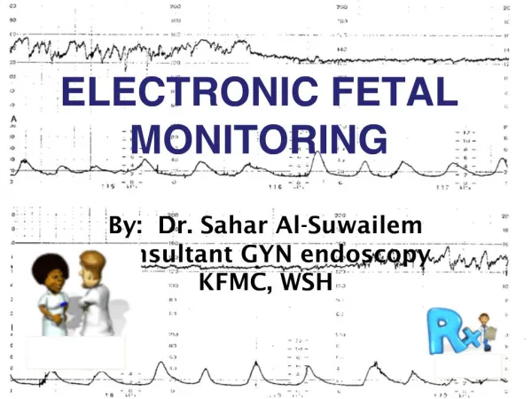
ELECTRONIC FETAL MONITORING (EFM) / CARDIOTOCOGRAPHY(CTG). Dr Rehana Raja King Khalid University Abha, KSA
Format • History • The methods available • Basic physiology • Indications • Features of CTG – Normal & Abnormal • Management of abnormal CTG • Fetal Blood Sampling • The future?
HISTORY • 1876 – Pinnarddesigned Pinnardsstethoscope • Early 1970s-Electronic fetal monitoring introduced in clinical practice • Early hopes were prevention of cerebral palsy and reduction of perinatal mortality • FHR patterns were thought to reflect hypoxia- fetal distress • EFM did NOT reduce Perinatal mortality but leads to an INCREASE of C-Sections
Fetal Monitoring in Labor: Two Acceptable Methods Electronic In “active” labor – by convention needs to be continuous Does not reduce perinatal mortality Increases c-section rates Variable interpretations Auscultatory - Pinnards Prescribed intervals Various devices but one recorded number Easy to interpret Intermittent Acceptable for “high” risk patients
Monitoring in an uncomplicatedpregnancy For a woman who is healthy and has had an otherwise uncomplicated pregnancy, intermittent auscultation should be offered and recommended in labour to monitor fetal wellbeing. In the active stages of labour, intermittent auscultation should occur after a contraction, for a minimum of 60 seconds, and at least: • every 15 minutes in the first stage • every 5 minutes in the second stage. Grade A Recommendation
Factors Necessary for Optimal Fetal Well-Being • Intact, functional maternal physiology • Intact, functional placenta • Intact, functional fetus
PROBLEMS with EFM • EFM does not improve perinatal mortality • Excess of operative deliveries ( ACOG 2009) • Interobserver and intraobserver variations in interpretation • Lack of consistency and standardization of definitions eg fetal distress—reassuring/non reassuring trace, pathological / suspicious • Lack of training/education and evaluation
In Practice a CTG is best regarded as a screening tool: • High negative predictive value • >98% of fetuses with a normal CTG will be OK • Poor positive predictive value • 50% of fetuses with an abnormal CTG will be hypoxic and acidotic but 50% will be OK • Therefore the CTG should always be interpreted in its clinical context • And backed by fetal blood sampling PRN
Indications for the use of continuous EFM
Selected High-Risk Indications for Continuous Monitoring of Fetal Heart Rate Maternal medical illness Gestational diabetes Hypertension Asthma Obstetric complications Multiple gestationPost-date gestationPrevious cesarean sectionIntrauterine growth restriction OligohydramniosPremature rupture of the membranesCongenital malformationsThird-trimester bleeding- AntepartumhaemorrhageOxytocin induction/augmentation of laborPreeclampsia Meconium stained liquor
Documentation The following should be recorded • woman’s name and MRN, • estimated gestational age, • clinical indications for performing the CTG • time and date • maternal pulse rate. • Signature with time and date • The outcome of the FHR pattern should be documented both on the CTG and in the woman’s medical records and signed by the doctor
BASICS • Speed of paper is usually 1cm per minute – hence I big square is 1 minute • The units used on the paper – 1 small square is 5 beats in the vertical axis • Sleeping cycle of fetus is 30 t0 40 mins – CTG should be done for atleast 20 to 30 mins- one can stimulate to awaken the baby like acoustic stimulation or a simple tap on the abdomen • CTG can be used in the antenatal period for fetal surveillance –Stress and non stress tests • Should NOT be done on Fetuses < 28 weeks
Baseline Heart Rate Short term variability Accelerations Decelerations Response to stimuli Contractions Fetal movements Others eg drugs egpethidine Features of a CTG
Baseline Fetal Heart Rate • Normal rate 110 to 150 bpm at term • Faster in early pregnancy • Below 100 = baseline bradycardia • Below 80 = severe bradycardia • Tachycardia > 160 bpm • Tachycardia if mother has fever
BRADYCARDIA 22
Hypoxia ChorioamnionitisMaternal fever B-Mimetic drugsFetal anaemia,sepsis,ht failure,arrhythmias TACHYCARDIA 23
Short Term Variability orBeat to Beat Variability • Should be 10 to 25 beats • The most important feature of any CTG • Is a reflection of competing acceleratory and decelerating CNS influences on the fetal heart • Represents the best measure of CNS oxygenation • Will be affected by drugs • Will be reduced in the pre term fetus
REDUCED VARIABILITY Hypoxia Drugs Extreme prematurity Sleep CNS abno. 26
SINUSOIDAL Dr Mona Shroff www.obgyntoday.info 27
Sinusoidal pattern A regular oscillation of the baseline long-term variability resembling a sine wave. This smooth, undulating pattern, lasting at least 10 minutes, has a relatively fixed period of 3–5 cycles per minute and an amplitude of 5–15 bpm above and below the baseline. Baseline variability is absent Associated with - Severe chronic fetal anaemia Severe hypoxia & acidosis 28
Accelerations • Must be >15 bpm and >15 sec above baseline • Should be >2 per 15 min period • Always reassuring when present • May not occur when fetus is “sleeping” • Should occur in response to fetal movements or fetal stimulation • Non reactive periods usually do not exceed 45 min • >90 min and no accelerations is worrying
Decelerations • Early: mirrors the contraction • Typically occurs as the head enters the pelvis and is compressed, i.e. it is a vagal response • Late: Follows every contraction and exhibits a slow return to baseline • Is quite rare but is the response of a hypoxia • Variable: Show no relationship to contractions • Mild • Moderate • Severe • In practice many “decels” or “dips” are MIXED
DECCELERATIONS EARLY : Head compression LATE : Utero placental insufficiency VARIABLE : Cord compression Primary CNS dysfunction 32
EARLY 33
Early decelerations • Begin with head compression. • This reduction of cerebral blood flow leads to hypoxia and hypercapnia • Hypercapnia leads to hypertension with triggering of baroreceptors • Results in bradycardia mediated by parasympathetic nervous system (via the vagal nerve) • Fall in FHR is matched to rise in contraction strength • Not indicative of fetal compromise
LATE 36
Late Decelerations • Repetitive from one contraction to the next (3 or more) • Recovery to baseline is late, well after the end of the contraction • More ominous when associated with minimal variability & baseline • Reflects a change in placental ability to adequately meet fetal needs • May indicate the presence of fetal hypoxia and acidosis • Often signifies fetal decompensation
VARIABLE 38
Variable Decelerations • Repetitive or intermittent • Often mimic letters of the alphabet U V W M • Rapid sudden fall in FHR • Often rapid recovery • Reflect some degree of umbilical cord impingement • Often seen when liquor volume is
FHR evaluation Dr C Bravado ALSO • DR – determine the risk • C – contractions • Bra – baseline rate • V – variability • A – accelerations • D – decelerations • O – overall assessment (followed by a management plan)
Maternal Position Dehydration Infection Hypotension ?Vaginal exam/bedpan Vomiting/vasovagal Analgesia/Drugs Mechanical Poor quality CTG Maternal pulse Transducer site Fetal scalp electrode Oxytocics Prostaglandins Suspicious FHR Pattern: What should you do?
Pathological: What should I do? • Roll woman into left lateral position, give oxygen, iv fluids & continue CTG monitoring • Perform Fetal Blood Sampling • If pH 7.25 repeat within one hour if the FHR abnormality persists • If pH 7.21-7.24 repeat within 30mins or deliver if rapid fall since last FBS • If pH < 7.20 DELIVER immediately • Lactate 4.2 - 4.8DELIVER– brain injury begins at 6mmols or higher • All FBS should take into account previous pH, rate of progress & clinical information
And finally… For the electronic fetal monitoring to be effective, the test must be performed correctly, its results must then be interpreted satisfactorily and finally this interpretation must provide an appropriate response Room for newer methods?? DEFINITELY!!! THANK YOU