Download
1 / 24
240 likes | 535 Views
Procedures. Intermediate Format Temporomandibular Joint Arthroscopy. Objectives. Assess the related terminology and pathophysiology of the TMJ. Analyze the diagnostic interventions for a patient undergoing a _______________.

E N D
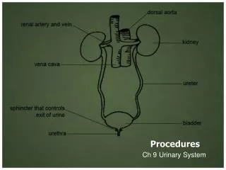
Procedures Intermediate Format Temporomandibular Joint Arthroscopy
Objectives • Assess the related terminology and pathophysiology of the TMJ. • Analyze the diagnostic interventions for a patient undergoing a _______________. • Plan the intraoperative course for a patient undergoing_____________. • Assemble supplies, equipment, and instrumentation needed for the procedure.
Objectives • Choose the appropriate patient position • Identify the incision used for the procedure • Analyze the procedural steps for TMJ Arthroscopy. • Describe the care of the specimen
Terms and Definitions • Bruxism • Malocclusion
Definition/Purpose of Procedure Temporomandibular Joint (TMJ) Disorder occurs when the muscles used in chewing and the joints of the jaw fail to work in combination with each other.
Pathophysiology Causes: Bruxism, Malocclusion, Arthritis, Trauma
Pathophysiology Signs & Symptoms Pain, clicking, limited range-of-motion, spasms, asymmetry
Diagnosis Linear CT and MRI
Treatment • 5-10 % dx w/TMJ Dysfunction fail to have relief of medical tx, and require surgery • Antiinflammatories, soft diet, hot compresses, muscle relaxants • >2 weeks: intraoral occlusion splints, med tx • Recurrent or chronic: permanent dental correction
Surgical Intervention:Special Considerations • Patient Factors • Outpatient • H& P, Blood chemistries, CBC, PT, PTT, U/A, serum HCG, Chest x-ray or ECG as appropriate • Room Set-up • X-rays in room
Surgical Intervention: Positioning • Position during procedure • Supine w/head donut pillow, tuck arms to side • Supplies and equipment • Arm sleds, headring pillow • Special considerations: high risk areas • Elbows—ulnar nerves • Prep • Shave preauricular area • Cotton to ears to prevent pooling of povidone-iodine & caution w/eyes; entire facial area prepped from hairline, down to shoulder, and laterally to include mouth and chin
Surgical Intervention: Special Considerations/Incision • Special considerations • Nasal intubation • Prophylactic antibiotics & steriods • State/Describe incision • Small stab incision w/# 11 before trocar is introduced at superior joint space
Surgical Intervention: Supplies • General: basic pack drape and split head sheet, gowns & gloves, towels, basin set, prep set, sterile adhesive wound drape, irrigation pouch, skin marker, raytex, • Specific • Suture & Blades (# 11) • Medications on field (name & purpose) • Catheters & Drains: n/a • Drapes: head turban for initial drape; pad pt forehead with a folded towel; plastic adhesive wound drape to cover ET tube and mouth; split sheet and large sheet for body drape, (laser: 4 wet towels around pt’s face; moistened cotton in external auditory canals, irrigation collection pouch at base of ear and TMJ)
Surgical Intervention: Supplies cont’d • 2 60 mL syringes • 4 10 mL syringes • 1 1-mL syringe • Needles: 18 g, 21 g, 25 g • Skin stapler • Eye pads • Sterile water and saline • 1000 mL Lactated Ringers for irrigation • 30 in extension tubing • Stopcock
Surgical Intervention: Instruments • General: suction, Lactated Ringer’s IV bag for irrigation, marking pen • Specific • TMJ instrument set • 0 degree arthroscope • 30-degree arthroscope • 70-degree arthroscope • Cannulas • Sharp & dull obturators • Light cord, camera & cord, small joint rotary shaver
Surgical Intervention: Equipment • General: suction system • Specific • Monitor/light source/camera tower, shaver control unit, IV pole for irrigant • Fluid infusion system • Bipolar ESU • Holmium laser
Surgical Intervention: Procedure Steps • Irrigation solution is injected into the joint space to distend the capsule • LR solution is preloaded in syringe w/needle attached. • After small stab incision is placed, surgeon inserts a sheath w/sharp obturator into superior joint space. After space is entered, the sharp is replaced with a dull obturator to further direct the sheath into the joint without damaging the intraarticular tissue or adjacent neurovascular structures. • #11 blade with # 7 handle will be ready • Trocar/cannula is preassembled. Expect trocor to be returned. Be prepared to assist with connections of video/light cord connections.
Surgical Intervention: Procedure Steps • Irrigation is infused into the joint • LR solution is connected to the cannua via extension tubing • Joint is examined • Prepare to operate remote control for still photos • If functional surgery is needed, a second stab wound is made • Pass skin knife. Prepare additional equipment (probe, shaver, grasper) • Final visual inspection is performed • Additional photos may be taken
Surgical Intervention: Procedure Steps • Cannuale are removed and excess fluid removed • Prepare for closure; count • Wound is closed and dressing placed • Pass suture; prepare dressings, reorganize equipment & supplies if procedure is bilateral • Steps may be repeated contralaterally • Repeat steps
Counts • Initial: sponges and sharps • First closing • Final closing • Sponges • Sharps
Specimen & Care • Identified as n/a or as specified (eg chondromalacia) • Handled: routine, etc.
Postop Considerations • Immediate • ROM of jaw limited • Suction and Emergency airway supplies readily available; Elevate HOB 30 degrees • Ice for pain and swelling • Liquid or soft diet for several days • Prognosis: good—may recur if behaviors not resolved; PT may begin in 24-48 hrs post-op. • Complications: hemorrhage, infection, recurrence • Joint damage, destruction of middle ear ossicles, perforation into middle cranial fossa, injury to auriculotemporal nerve
Resources • www.healthscout.com • STST pp. 646-647, Procedure 18-7 • www.dentaljournal.com/article 6 • Rodau; Baker-Gill, Levin; “Arthroscopic Temporomandibular Joint Surgery”, AORNJournal Nov 1993, 58: 5.