Download
1 / 100
1.04k likes | 1.18k Views
Learn about developmental dysplasia of the hip (DDH) and other hip conditions in children, their causes, examination methods, diagnostic tests, and treatment options. Early detection and proper management are crucial for successful outcomes.

E N D
Common Pediatric Hip Problem Dr.Kholoud Al-Zain Assistant Professor Consultant, Pediatric Orthopedic Surgeon Dec 2016 Acknowledgement: Dr.Abdalmonem Alsiddiky Dr.Khalid Bakarman Prof. M. Zamzam
Common Pediatric Hip problems • DDH • SCFE • Perth's
Nomenclature • CDH : Congenital Dislocation of the Hip • DDH : Developmental Dysplasia of the Hip
Pediatric Hips Dislocation • Types: • Idiopathic isolated pathology • Teratologic: • Neurologic as: patient with C.P or MMC • Muscular as: Arthrogryposis • Syndromatic as: Larsen syndrome • Miscellaneous: • Complication to hip septic arthritis • Traumatic
Pediatric Hips Dislocation • Note delivery in its self (OBGY Dr.) does not dislocate a hip • DDH occurs in the 3ed trimester • Teratologic usually in the 1st trimester
Normal pelvis Adult Child Femoral head ossific nucleus Growth plates
DDH • The pathology is of 2 components: • Femoral head position • Acetabular development
Normal hip Dislocated hip 1) Femoral Head Position Superior displacement Femoral head lateralization
Normal hip Dislocated hip 2) Acetabular Development Acetabular dysplasia
Patterns of Disease • Dislocated • Dislocatable • Subluxate • Acetabular dysplasia (A.D)
Causes (multi factorial) Unknown • Hormonal • Relaxin, oxytocin • Familial • Lig.laxity diseases • Genetics • F 4-6x > M • Twins 40% • Mechanical • Pre natal • Post natal
Mechanical Causes • Pre-natal: • Breach • Oligohydrominus • Primigravida • Twins • Post-natal swaddling , strapping
Infants at Risk • Parents who are relatives (consanguinity) • Positive family history: 10X • 1st child • Breach presentation: 5-10 X • Oligohydrominus • Twins: 40% • A baby girl: 4-6 X • Torticollis: CDH in 10-20% of cases • Foot deformities: • Calcaneo-valgus • Metatarsus adductus • Knee deformities: • hyperextension and dislocation
DDH • When risk factors are present the infant should be reviewed: • Clinically • Radiologically
Examination • The infant should be: • Quiet • Comfortable
DDH • Look: • External rotation • Lateralized contour • Shortening • Asymmetrical skin folds • Anterior • Posterior
DDH • Move • Limited abduction
DDH • Special test (depending on the age): • Galiazzi sign • Ortolani, Barlow test only till 4-6 m of age • Hamstring Stretch test • Trendelenburg sign older comprehending child • Limping: • Unilateral one sided limping • Bilateral waddling gait (Trendelenburg gait)
Limb Length Inequality • Clinical measures of discrepancy: • Measuring tape • Giliazi test
DDH- Investigations • 3w -3m U/S • > 3months XR pelvis (AP + abduction) • > 5-6m: • More reliable • Is when ossification centers normally appears • If delayed or did not appear it’s one of the signs of DDH
DDH- Radiology Acetabular Index Perpendicular Line Horizontal Line Shenton's Line
Treatment - Aims • A concentrically, reduced, stable, painless, mobile hip joint: • Obtain concentric reduction • Maintain concentric reduction • In a non-traumatic fashion • Without disrupting the blood supply to femoral head • Parents education about inheritance That is why: Refer to pediatric orthopedic surgeon
DDH- Treatment • Method depends on age • The earlier started: • Its easier • Better the results (higher remodeling potential) • Treatment is mainly non-operative • Should be detected EARLY • Either surgical or non-surgical
Treatment • Birth – 6m • In OPD: reduce + maintain with Pavlik harness or hip spica (H.S) • 6-12 m: • GA + closed (? Open) reduction + maintain with H.S • 12 - 18 m: • GA + open reduction + maintain with H.S 6w, then B.S cast for months • 18 – 24 m: • GA + open reduction + acetabuloplasty + H.S 6w, then B.S cast 6w • 2-8 years: • GA + open reduction + acetabuloplasty + femoral shortening + H.S 6w, B.S 4-6w • Above 8 years: • GA +open reduction + acetabuloplasty (advanced) + femoral shortening + H.S
Pavlik Harness • Maximum to start it is 6m of age, if older use other method • This is to achieve stable reduction • It’s a dynamic splint • Is kept on for 6w continuous, then use a rigid abduction splint
Abduction splint • It’s a rigid splint • This is to: • Maintain the reduction, • And wait for improvement of the acetabular cover to be: • A.I < 30° • & with concavity
Normal Hip Arthrogram Acetabular cartilage Concentrically reduced femoral head
Hip Arthrogram Guided Reduction Dislocate view Reduced view
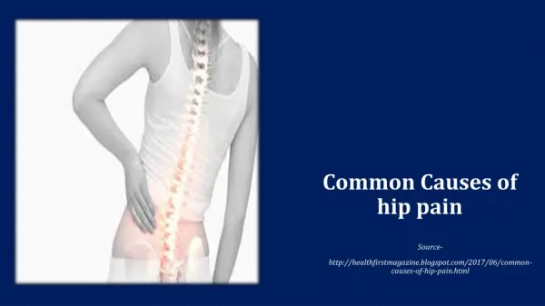
DDH • Late complications if not treated: • Severe pain (hip area, back) • LLD (leg length discrepancy) • Pelvic inequality (tilt) • Early hip arthritis • Early Lumbar spine degeneration • Secondary scoliosis
SCFE • Slipped Capital Femoral Epiphysis • At the level of physis • As if it is a Salter-Harris fracture, type-1 • So it is an emergency
SCFE- Top View Anterior slippage
SCFE • Types: • Radiological: • Acute < 3w • Chronic > 3w, can see start of callus formation • Acute on chronic • Clinical: • Unstable can not weight bear on that limb • Stable can put some weight (walk) • When it’s acute or unstable urgent surgery
SCFE • Causes (multifactorial): • Unknown • Hormonal: • Hypothyroid • Abnormal G.H • Hypogonadisum • Metabolic Chronic renal failure • Mechanical (obesity) • Trauma