Download
1 / 24
240 likes | 278 Views
Learn about the structure, dynamics, and functions of microfilaments (actin filaments) and intermediate filaments in cell motility, anchoring membrane proteins, and providing structural support. Understand the assembly processes, polarity, and roles in cell functions like anchorage, movement, and locomotion. Explore the unique characteristics, subunit composition, and mechanical support of intermediate filaments. Discover diseases linked to IF defects like epidermolysis bullosa and Alzheimer's.

E N D
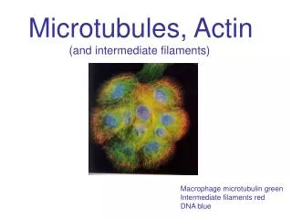
Microfilaments and Intermediate FilamentsPresented by: Leslie Hargis
Cytoskeleton- Cytoplasmic system of fibers, critical to cell motility • 3 Types: • Microfilaments (Actin Filaments) • Intermediate Filaments • Microtubules
Microfilaments and Intermediate Filaments • Both are usually attached to plasma membrane proteins • They form a skeleton that helps support the plasma membrane
Actin Filaments • Thinner, shorter and more flexible than microtubules • Contains G-actin, and F-actin • Actin- most abundant intracellular protein in most Eukaryotic cells • Comprises 10% by weight of total cell protein in muscle cells, 1-5% in non-muscle cells
Actin polymerization requires K+, Mg 2+, ATP, and Calcium • Here is an animation of the sliding of a single myosin molecule along an actin filament • www.sci.sdsu.edu/movies/actin_myosin_gif.html
G-actin and F-actin • G-actin: exists as a globular monomer • F-actin: is a helical filamentous polymer of G-actin subunits all oriented in the same direction • Microfilaments in a cell are constantly shrinking or growing in length • Bundles and meshworks of microfilaments are forming and dissolving continuously
Dynamics of Actin Assembly • Nucleation- G-actin clumps into short, unstable oligomers, 3-4 subunits long, and acts as a stable seed or nucleus. • Elongation phase- The nucleus rapidly increases in length by the addition of actin monomers to both of its ends.
Steady-State The ends of actin filaments are in a steady state with monomeric G-actin. After their incorporation into a filament, subunits slowly hydrolyze ATP and become stable F-actin. Dynamics of Actin Assembly
Dynamics of Actin Assembly • All subunits in an actin filament point toward the same direction of the filament • Exhibits polarity-actin subunit exposed to the surrounding solution is the (-) end • The cleft that has contact with the neighboring actin subunit that is not exposed is the (+) end • Actin filaments grow faster at the (+) end than the (-) end
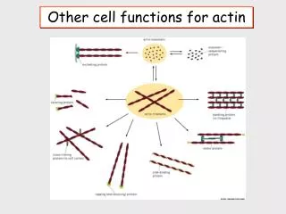
Actin Filaments participate in a variety of cell functions: • Anchorage and movement of membrane proteins- • filaments are distributed in 3-dimensional networks throughout the cell • used as anchors with in specialized cell junctions
Actin Filaments participate in a variety of cell functions: • Formation of the structural core of microvilli • On epithelial cells, help maintain shape of the cell surface
Actin Filaments participate in a variety of cell functions: • Locomotion of the cells • Achieved by the force exerted by actin filaments by polymerization at their growing ends • Used in many migrating cells, particularly on transformed cells of invasive tumors • Cells extend processes from their surface by pushing the plasma membrane ahead of the growing actin filaments
Functions and structure of intermediate filaments distinguish them from other cytoskeletal fibers
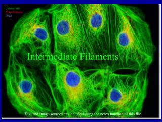
Intermediate Filaments (IF) • Found in most animals but not in plants and fungi • Smaller than microtubules but larger than microfilaments • Subunits are a-helical rods that assemble into ropelike filaments • Unlike microfilaments, IF’s don’t contribute to cell motility
Intermediate Filaments (IF) • Provides mechanical support for the plasma membrane where it comes in contact with other cells or with the extracellular matrix • Extremely stable- even after extraction with solutions containing detergents and high concentrations of salts
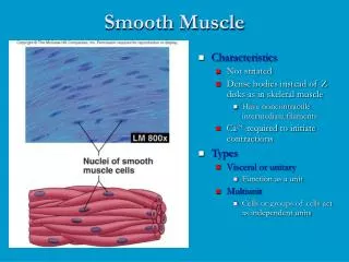
IF’s are broken down into 4 groups: • Lamins- found in the nucleus • Keratins (cytokeratins)- in the epithelia • Acidic or basic • “hard” epithelial tissues- nails, hair, wool
IF’s are broken down into 4 groups: • Type III IF proteins (Vimentin)- most abundant type • In leukocytes, blood vessel endothelial cells, mesenchymal cells • Neurofilaments- neuronal axons • Extend from the cell body into the ends of axons and dendrites • Provides structual support
Intermediate Filament Assembly • Assembled from a pair of helical monomers that twist around each other to form coiled-coil dimers • Then 2 coiled-coil dimers twist around each other to make a tetramer of 2 coiled-coil dimers • This forms the non-polarized unit of the IF’s (unlike microfilaments that are polarized)
Diseases caused by defects in the IF • Epidermolysis bullosa simplex • Blisters form due to lack of normal bundles of keratin filaments • Alzheimer’s disease • Caused by changes in the neurofilaments with in brain • Alcoholic liver cirrhosis • Accumulation of keratin filaments forming inclusions called mallory bodies in liver