Download
1 / 97
1.12k likes | 1.73k Views
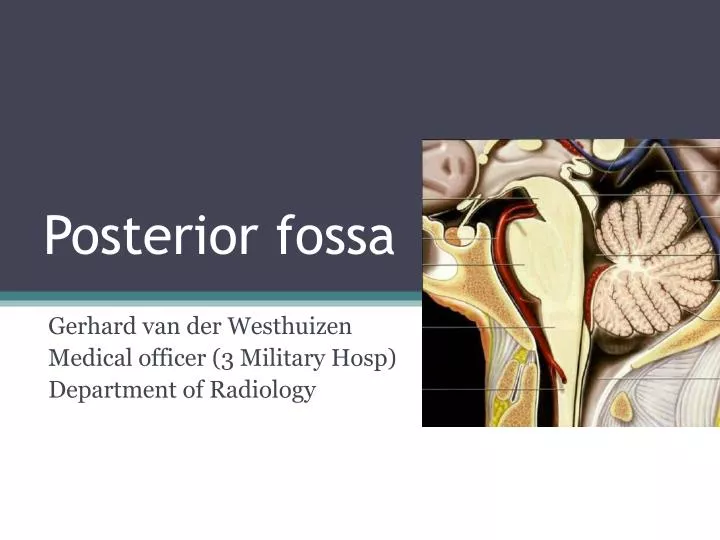
Posterior fossa. Gerhard van der Westhuizen Medical officer (3 Military Hosp) Department of Radiology. Posterior fossa - Outline. Calvarium Posterior skull base Brainstem anteriorly Midbrain, pons and medulla Cerebellum posteriorly 2 Hemispheres and midline vermis Divided into:

E N D
Posterior fossa Gerhard van derWesthuizen Medical officer (3 Military Hosp) Department of Radiology
Posterior fossa - Outline • Calvarium • Posterior skull base • Brainstem anteriorly • Midbrain, pons and medulla • Cerebellum posteriorly • 2 Hemispheres and midline vermis • Divided into: • Mesencephalon (midbrain) • Rhomboencephalon (pons, medulla and cerebellum) • Cerebral aquaduct and fourth ventricle • CSF cisterns containing vertebrobasilar arteries and veins
Posterior skull base • Formed by posterior temporal and occipital bones • Anterior - Dorsum sellae medially, petrous ridges laterally • Posterior - Groove for transverse sinus on occipital bone • Transmits CN 7-12, medulla oblangata and jugular veins • Multiple foramina and fissures
Posterior skull base -Foramina • Internal acoustic meatus • Porusacusticus – CN VII & VIII, labyrinthine artery • Jugular foramen • Pars nervosa - anteromedial • CN IX, Jacobson’s nerve and inferior petrosal sinus • Pars vascularis - posterolateral • Jugular bulb, CN X & XI, Arnold’s nerve, posterior meningealartery, meningeal branch of ascending pharyngeal artery • Hypoglossal canal • CN XII • Stylomastoid foramen • CN VII • Foramen magnum • Medulla oblangata, CN XI and vertebral arteries
Brainstem • Midbrain • Connects pons and cerebellum with forebrain • Pons • Relays information from brain to cerebellum • Medulla • Relays information from spinal cord to brain
Midbrain (Mesenchephalon) • “Butterfly-shaped”, passes through tentoriumcerebelli • 3 Main parts: • Cerebral peduncles • White matter tracts - Corticospinal, corticobulbar & corticopontine tracts • Tegmentum • CN nuclei: III– Level of superior colliculus; IV – Level of inferior colliculus Accessory oculomotor (Edinger-Westphal) • Gray matter nuclei • Substantianigra- Motor planning, eye movement, reward seeking, learning and addiction • Red nucleus – Relay and control centre of cortiomotor impulses. • Periaquaductalgray matter – Pain and defensive behaviour • White matter tracts • Spinothalamic • Medial and lateral lemniscusSomatosensory • Medial longitudinal fasciculus – Vestibulo-ocular and optokinetic reflexes
Midbrain • Tectum • Superior colliculus( visual pathway) • Inferior colliculus(auditory pathway) • Cerebral aquaduct passes between tectum and tegmentum • CSF cisterns associated with midbrain • Ambient – Lateral, CN IV • Quadrigeminal – Posterior, CN IV • Interpeduncular – Anterior, CN III. • Connections: • Superior – Cerebral hemispheres, basal ganglia and thalami • Posterior – Cerebellum viasuperior cerebellar peduncle (brachium conjuntivum) • Inferior – Pons • Blood supply via vertebrobasilar circulation • Perforating branches of basilar, SCA, PCA.
Pons • Relays info from brain to cerebellum. • Middle cerebellar peduncle – Brachium pontis • Bulbous midportion of brainstem • Two main parts: • Ventral pons – White matter tracts continuous with cerebral peduncles and medullary pyramids. • Dorsal tegmentum– CN nuclei, gray matter nuclei and white matter tracts. Continuation of midbrain tegmentum superiorly and medullarytegmentum inferiorly.
Pons • Tranverse fibres make up bulk • Dorsal surface forms rostral half of 4th ventricle. • Adjacent CSF cisterns: • Prepontine – CN V & VI • CP angle – CN VII & VIII • Blood supply • Medial branches SCA, perforating branches of basilar artery, thalamoperforator arteries.
Pons • CN nuclei: • V – Throughout brainstem and upper cord. • Bulk of motor and sensory in pons. • Enters and exits at level of midlateralpons • VI– In pontinetegmentum, near midline, anterior to fourth ventricle. • Exits anterior at ponto-medullary junction • VII– Ventrolateral aspect of pons • Motor, superior salivatory, solitary tract • Exits laterally at ponto-medullaryjunction VIII – Vestibular along floor of 4thventricle Cochlear on lateral surface of inferior cerebellar peduncle • Exits at ponto-medullary junction, posterior to VII
Medulla • Caudal part of brainstem composed of gray matter formations, CN nuclei IX – XII and white matter tracts. • Between pons and spinal cord. • 4th ventricle and cerebellum posteriorly • Connected to cerebellum via inferior cerebellar peduncle (restiform body). • 2 Main parts: • Ventral – olive and pyramidal tract • Dorsal tegmentum – CN nuclei and white matter tracts
Medulla • Ventral medulla: • Pyramid • Paired; anterior surface; midline ventral median fissure • Ipsilateralcorticospinal tracts prior to decussation • Olive • Lateral to pyramids, venterolateralsulcus (pre-olivary) and posterolateralsulcus (post-olivary) • Inferior olivary complex of nuclei
Medulla • Dorsal tegmentum: • Multiple white matter tracts. • Gracile and cuneate tubercles • Lower aspect of dorsal medulla • Nuclei gracilis(medial) ; cuneatus (lateral) • Fourth ventricle terminates in caudal medulla. • Blood supply: • Distal vertebral arteries • PICA • Anterior spinal artery