Download
1 / 53
540 likes | 852 Views
INNATE IMMUNITY BASIC MECHANISMS. determines „context“ for specific immunity. fylogenetically conserved. relationship to other systems. INNATE IMMUNITY. identification of „danger“ patterns. immediate reactivity. ontogenically conserved. natural cytotoxicity. interferons.

E N D
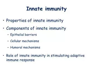
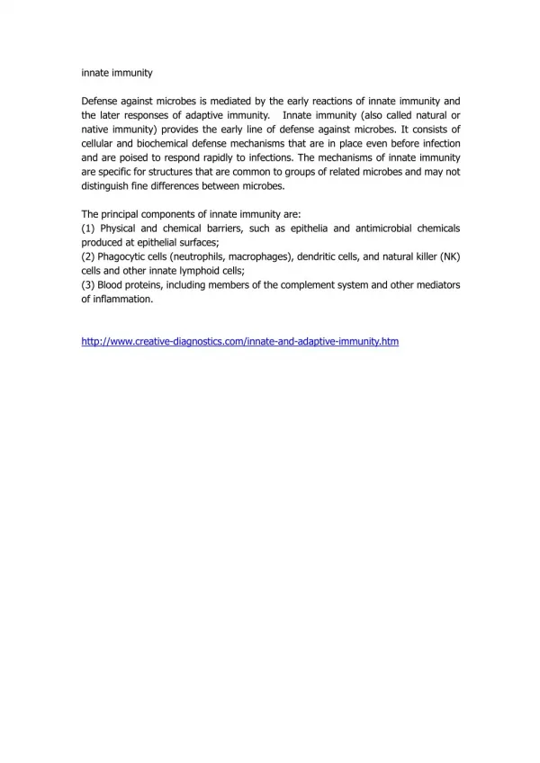
INNATE IMMUNITY BASIC MECHANISMS
determines „context“ for specific immunity fylogenetically conserved relationship to other systems INNATE IMMUNITY identification of „danger“ patterns immediate reactivity ontogenically conserved
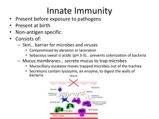
natural cytotoxicity interferons complement system INNATE IMMUNITY phagocytosis macrophages dendritic cells granulocytes
identification of „danger“ patterns processing and presentation of antigens exogenous endogenous dendritic cells macrophages context for specific recognition
L L L L L L APC dendritic cell macrophage MBL MASP FcR IL-10 TGF context for specific recognition Tr reactivity IL-1, IL-12, TNF costimulation TH1 reactivity IDENTIFICATION OF PATHOGEN ASSOCIATED MOLECULAR PATERNS (PAMPS) lipoteichoic acide peptidoglycan, lipoprotein sacharide polyanions pathogenicity island phosphoryl cholin TLR2 LPS TLR4 CpG TLR9 pentraxins (CRP) LPS LBP SCAVENGER RECEPTOR LECTIN cell activation IL-1R CD14 IL-1R LPS IL-1R MyD88 DD DD TRAF6 NIK CR C3b IRAK I-B I-B NF-B ubiquitination degradation NF-B cell activation Y activation of complement by lectin TRANSCRIPTION NF-B NF-B IL-1 IL-12 IL-6 IL-8
immunopathology tumor infection dendritic cells macrophages killing of intracellular microbes ACTIVATION CYTOKINES TH1 TH2 polarisation stimulation of hematopoiesis pluripotent proinflammatory chemokines INFLAMMATION
ROLE OF DENDRITIC CELL IN INNATE IMMUNITY capture and processing of antigens in tissues presentation of antigens to T cells DENDRITIC CELL migration into lymphatic nodes
ROLE OF GRANULOCYTES IN INNATE IMMUNITY GRANULOCYTES stimuli hematopoiesis endo- exo- activation professional phagocytes cytokines adhesion to endothelia inflammation diapedesis killing and destroying of microbes
ROLE OF INTERFERONS IN INNATE IMMUNITY most important humoral factors of innate immunity inducible induced by viral (microbial) agents, nucleic acids active after binding to cell receptors protect against viral infections immunomodulatory activity
II nd class INF (TH1) I st class INF ,, immunomodulatory activities antiviral activity proinflammatory activities INTERFERONS antimicrobial activity cytotoxicity expression HLA II T ly NK antiproliferative activity anticancer activity
ANTIVIRAL ACTION OF INTERFERONS infected cell interferons Jak STAT uninfected cell interference with viral proteosynthesis cleveage of viral nucleic acid VIRAL UNRESPONSIVNESS
vmRNA vmRNA vdsRNA 2´5´oligo- adenylats ATP 2´-5´-oligo- adenylat- synthetasis induction of 2´5´oligoadenylat- synthetasis ANTIVIRAL ACTION OF INTERFERONS inhibition of viral proteosyntesis degradation of mRNA RIBOSOM mRNA U A A U A U G ElF4 ElF3 ElF2 UA C P PKR active P ElF4 ElF2 RNAáza L aktivní P GTP ElF2 Met dsRNA + ATP Met ElF3 PKR inactive RNAáza L inaktivní induction of protein kinasis (PKR) IFN receptor IFN
COMPLEMENT SYSTEM humoral factor of innate immunity COMPLEMENT SYSTEM synthesis hepatocytes 30 plasma and membrane proteins macrophages amplification cascade
FUNCTION OF COMPLEMENT SYSTEM anafylactic cytolytic elimination of immune complexes opsoniziing proinflammatory chemotactic
NOMENCLATURE OF COMPLEMENT SYSTEM components of complement system: C1 … C9 factors of complement system: designed by capital letters (P, B, D, H) enzymatically active complex: (C4b2a) fragments of complement system: small letters (a, b) fragment b: enzymatically active (C3b) fragment a: anafylactically active (C3a)
ACTIVATION OF COMPLEMENT SYSTEM enhanced activation cascade principle - proactivation conditions - active enzyme (serine protease) - cleveage of the next component - active enzyme formation tight regulation of activation on several levels C3, C5 convertases formation are crucial steps of activation
AMPLIFICATION CASCADE stimuli (microbial, contact activation, coagulation) resting component (factor) of complement system proteolytically active activation (proteolytic cleveage) proinflammatory anafylactic, chemoattractive activities
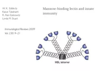
CLASSICAL PATHWAY OF ACTIVATION OF COMPLEMENT SYSTEME starts on complex AgAb (IgM, IgG1, IgG3) aggregates of immunoglobulins C3 convertase: C4b2a C5 convertase: C4b2a3b LECTIN PATHWAY OF ACTIVATION OF COMPLEMENT SYSTEME specific manose sugar residues are identified by MBP (manose binding protein - lectin) MASP serine protease is consequently activated
ALTERNATIVE PATHWAY OF ACTIVATION OF COMPLEMENT SYSTEME spontaneous C3 component activation in the presence of H2O activated C3b is rapidly deactivated in the absence of proactivation conditions C3b is not deactivated in the presence of proactivation molecules. proactivation molecules: of either microbial origin (surfaces, LPS, teichoic acid) or agregates of immunoglobulins or arteficial surfaces C3 convertases: PC3bBb C5 convertases: PC3bBbC3b
TERMINAL STEPS OF ACTIVATION OF COMPLEMENT SYSTEME alternative and classical pathways of complement activation are separated C5 activation level solid phase is necessary for C3 and C5 convertases formation small fragments (C5a, C4a) with anafylactic and chemotactic activity are formed
ACTIVATION OF COMPLEMENT lectine pathway alternative pathway classical pathway C1q, r, s MBP MASP C3 convertases of either classical or alternative pathway C5 convertases of either classical or alternative pathway complex MAC lysis
TERMINAL PHASE OF COMPLEMENT ACTIVATION unified for both pathways nonenzymatic, starts from membrane bound C5b component conformational changes of complement components membrane attack complex (MAC) formation cell membrane perforation
MEMBRANE REGULATION OF COMPLEMENT ACTIVATION the expression of membrane regulatory proteins which prevent MAC complex formation protectin (CD59) decay accelerating factor (DAF. CD55)
RECEPTORS FOR COMPONENTS OF COMPLEMENT expressed on T and B cells (regulation of their functions) expressed on phagocytes (opsonisation) expressed on erythrocytes (immune complexes elimination) CD classified (CD 35, CD21, CD11)
COMPLEMENT SYSTEM - CLINICAL REMARKS INHERITED DEFECTS are very rare early components deficiency: prone to immunopathological diseases (SLE) C1 esterase deficiency: hereditary angioedema late components deficiency: susceptibility to meningoccocal infections
COMPLEMENT SYSTEM - CLINICAL REMARKS ACQUIRED DEFECTS frequent in systemic tissue disorders (SLE) enhanced complement activation in dialysis, extracorporal circuits deregulated activation of complement in patients with systemic inflammatory response syndrome (SIRS)
COMPLEMENT SYSTEM - LABORATORYREMARKS all components, factors and activation fragments of complement could be determined immunochemically C3, C4 components are acute phase proteins evaluation of activation of complement in vitro (CH50 level)
ingestion of microbes PHAGOCYTOSIS cell factor of innate immunity macrophages presentation of antigens dendritic cells granulocytes PHAGOCYTOSIS is the ability of specialised cells: to engulf to kill to destroy microbes and foreign particules source of cytokines
MONOCYTE - MACROPHAGES: • long-living tissue cells • principal source of pluripotent • proinflammatory cytokines • processing and presentation of antigens to T cells • immunoregulatory functions- killing • of intracellular microbes (M. tuberculosis)
GRANULOCYTES: professional phagocytes short-lived blood cells rapid turnover (G-CSF) highly glycosylated surface molecule CD15 cytoplasmic granules
PHAGOCYTOSIS: activation adhesion chemotaxis ingestion killing and destruction ACTIVATION RESULTS IN: increased genes transcription increased metabolic activity changes in cell shape changes in cytoskeleton changes in surface molecules
ACTIVATION OF PHAGOCYTES ENDOGENOUS: EXOGENOUS: proinflammatory cytokines C3a C5a PAF LT bacterial components LPS, CpG, „danger patterns“ (PAMP) chemokines CSF
ACTIVATION OF PHAGOCYTES changes in membrane molecules expression changes in cytoskelet changes in metabolism
ADHESION activated granulocytes activated endotelial cells DIAPEDESIS
A D H E S I O N • firm adherence of activated neutrophils • to activated endothelial cells • is prerequisited for diapedesis • mediated through the specific interactions • between adhesion molecules and their ligands • expression of adhesion molecules is inducibile • (under cytokine control) • selective adhesion together with chemokine signalling • is responsible for selective migration • of different leukocyte populations
ADHESION MOLECULES families are subdivided according to their structural characteristics FAMILY OF IMMUNOGLOBULINS immunoglobuline domain ICAM-1, 2, 3 VCAM-1 many others
FAMILY OF INTEGRINS: heterodimers (, chains) common type of chain determinates subfamily of integrins 1(CD29) integrins: - VLA molecules - ligands are molecules of ECM (collagen) 2(CD18) integrins: - leukocyte integrins - LFA-1 (CD11a/CD18) - ligand is ICAM-1 (CD54) 3integrins: - cytoadhesins - platelets
FAMILY OF SELECTINS terminal part of molecule is lectin-like ligands are sugars absent on resting cells rapidly mobilized from intracellular stores E-selectin (CD62E): - activated endothelia - ligand is a sugar on CD15 molecule P-selectin (CD62P): - activated platelets - activated endothelia - ligands are various glycosylated molecules (PSGL-1, CD 162) L-selectin (CD62L): - leukocytes - ligand is CD34 molecule on endothelium
CADHERINS IMMUNOGLOBULINS ICAM-1, 2, 3, VCAM-1 SELECTINS E, P, L - selectins sugar ligands ADHESION MOLECULES INTEGRINS heterodimers 7 homing 1 integrins (VLA) ECM binding 3 integrins cytoadhesins 2 integrins leukocytic (LFA-1)
ADHESION MOLECULES proinflammatory cytokines, chemokines inducibile macrophages dendritic cells immuno- pathology infection tumor
1ST STEP OF ADHESION: rapid upregulation (minutes) of selectin molecules on endothelial cells by the action of proinflammatory cytokines interaction between E-selectin and CD15 weak interaction (tethering) rolling of leukocytes on activated endothelia
IST STEP OF ADHESION, SELECTINE MEDIATED collagens IMMUNOPATHOLOGY a1 b1 fibronectin a1 b1 INFECTION proinflammatory proadhesive signals a3 b1 MALIGNANCY MACROPHAGE CD62E IL-1 TNF CD34 CD62P E-CADHERIN CHEMOATTRACTANTS CD15 CD62L PECAM-1 PSGL-1 ENDOTHEL GRANULOCYTE rolling
2ND STEP OF ADHESION firm adhesion interaction between LFA-1 and ICAM-1 signaling: outside-in inside-out cell spreading 3RD STEP OF ADHESION diapedesis into tissues
4 b1 b2 II ND STEP OF ADHESION A C T I V A TION C-X-C CHEMOKINES ENDOTHELIA MACROPHAGES C-C CHEMOKINES ICAM-1 VCAM-1 VLA-4 LFA-1 ICAM-1 DIAPEDESIS EOSINOPHIL LFA-1 b2 NEUTROPHIL 3CYTOADHESINS FIBRINOGEN vWF
non-random movement of granulocytes in the gradient of chemoattractants endogennous chemoattractants complement C5a, C3a CHEMOTAXIS chemokines exogennous chemoattractants CXC CC ELR+ ELR- microbial products
CHEMOTAXIS serpentin receptors for chemoattractants diapedesis binding of chemokines on proteoglykans and erythrocytes, neutralisation of biological activity of chemokines ECM degradation MMP, TIMP
INGESTION: close contact with microbe is prerequisited opsonins: specific antibodies complement components CRP surface receptors for: Fc fragment of immunoglobulins C3b component of complement lectins sugars interactions phagosome is fused with cytoplasmic granules
GRANULOCYTE BACTERIA Ig FcR Ag A C T I V A T I O N S-S C3b ITAM CR sacharide lectin lectinophagocytosis INGESTION
INTRACELLULAR KILLING AND DEGRADATION O2 independent O2 dependent defensins NADPH oxidase
NEUTROPHIL GRANULOCYTE - GRANULE CONTENT primary granule defensins MPO lysosyme BPI lactoferin cathepsin-G elastase proteinase-3 secondary granule cytochrome b558 integrins lysosyme lactoferin collagenase