Download
1 / 70
810 likes | 2.69k Views
ACUTE SURGICAL INFECTION. Non-Specific Acute Infection Specific Acute Infection . Non-Specific Acute Infection. Postoperative Wound Infection Cellulitis Erysipelas

E N D
ACUTE SURGICAL INFECTION Non-Specific Acute Infection Specific Acute Infection
Non-Specific Acute Infection • Postoperative Wound Infection • Cellulitis • Erysipelas • Boil (Furuncle) • Carbuncle • Hydradenitis Suppurativa • Acute Abscess • Acute Lymphangitis and Lymphadenitis • Bacteraemia and Septicaemia
Specific Acute Infections • Tetanus • Gas Gangrene • Necrotizing Fasciitis
Postoperative Wound Infections Are caused by the presence of contaminating microbes derived from : Endogenous OR Exogenous
Postoperative Wound Infection Predisposing Factors: # General 1- Poor general condition 2- Systemic disease 3- Drugs that cause immunosuppression
Postoperative Wound Infection # Local: 1- Poor blood supply 2- Poor surgical technique 3- Presence of foreign bodies 4- Nature of the operation 5- Defect in sterilization technique in the operating theatre
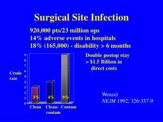
Types of surgical wounds Operative wounds are divided into three categories :1- Clean The risk of infection is 1-2%2- Clean contaminated The risk of infection 2-5%3- Contaminated The risk of infection is 5-30%
Pathology Acute inflammatory stage with local vasodilatation and infiltration by polymorph nuclear leucocytes. This is followed by suppuration with purulent discharge
Clinical Picture Wound infection usually appears between the fifth and tenth days postoperative * Fever * Pain in the wound Signs:_ swollen _tenderness _redness _fluctuant
Differential Diagnosis Other causes of postoperative fever • chest infection • DVT • UTI Other causes of wound swelling • heamatoma
Prophylaxis * Improve the defense mechanism * Control the predisposing factors * Prophylactic antibiotics * In bowel surgery, mechanical and chemical preparation of the bowel * Meticulous surgery * Operation in septic areas with heavily contaminated wounds should be left open
Treatment • Surgical drainage of the pus • Antibiotics in invasive infections • Look for hospital acquired infection
Cellulitis • Is an invasive non suppurative infection of the loose connective tissue • Organism : • streptococci [common] • staphylococci [occasionally] • mix
Clinical picture • The affected area is red,indurated,hot and painful • It spreads rapidly with ill defined edge • The skin may be the seat of blisters • Fever • Lymphangitis in the form of red streaks • No suppuration • In severe cases patches of skin necrosis with sloughing of subcutaneous tissues
Differetial Diagnosis • Contact allergy • Chemical inflammation • DVT
Treatment • Rest and elevation of the affected part • Antibiotic penicillin iv
Erysipelas Is a rapidly spreading non-suppurative inflammation of the lymphatics of the skin caused by a specific strain of hemolytic streptococci
Clinical Picture • Toxemia • Locally : similar to cellulitis,but there are the following differences: 1. The color of the skin is rose-pink 2. The edge is well defined 3. There may islets of inflammation beyond the spreading margin
Complications • 1. Facial erysipelas may lead to cavernous sinus thrombosis • 2. Septicemia • 3. Recurrent erysipelas may block the lymphatics leading to elephantiasis.
Treatment • Isolation • Similar to cellulitis
Boil [Furuncle] • Is a staphylococcal infection of a hair follicle or a sebaceous gland. • The common sites: face, neck and axilla. • Common in diabetics.
Clinical Picture A small painful indurated swelling which is - red - hot - and very tender
Treatment • 1.Antibiotics. • 2.Antiseptic.
Carbuncle • Is infective gangrene of the subcutaneous tissues usually secondary to infection by Staphylococcus aureus. • It is common in immunocompromised patients as in diabetics. • The common sites: face, nape of the neck, and the back
Pathology • Infection usually starts in a hair follicle • Extends to the subcutaneous fat where other hair follicles get the infection. • Multiple areas of necrosis and thrombosis of blood vessels occur. • Patches of skin undergo sloughing and separate from the underlying granulation tissue
Clinical Picture • There is usually sever toxemia. • Starts as a painful induration of the skin and subcutaneous tissues. • The skin is red. • Swelling its central part becomes soft. • Multiple areas of skin thin out and separate forming multiple sinuses.
Complications • Local spread of infection. • Pyaemia and septicemia. • Cavernous sinus thrombosis • Epidural abscess or meningitis
Treatment 1.Antibiotics. 2.culture and sensitivity of the discharge. 3.control of diabetes. 4.surgical excision of sloughs.
Hydradenitis Suppurativa Mixed staph. And streptococcal infection of the apocrine sweat glands, in the perineum or the axilla,produces multiple abscesses and pus discharging sinuses.
Treatment • Surgical drainage of abscesses. • Antiseptic and antifungal applications. • Surgical excision of the apocrine sweat-bearing skin following by skin grafting is essential.
Acute Abscess • It is a localized suppurative inflammation. • It is caused by pyogenic organisms. The commonest are staphylococci that produce a coagulase enzyme.
Pathogenesis The organism reach the tissues by : - direct access through wounds, scratches and abrasions. - local extension from an adjacent focus - lymphatic spread. - blood spread.
Pathology An abscess consists of three zones: 1- A central zone of coagulative necrosis 2- An intermediate zone of granulation tissue. 3- A peripheral zone of acute inflammation.
Sequlea • Resolution. • Pointing and rupture. • Spread infection – locally - by lymphatics or blood • Chronicity.
Clinical Picture • Locally :- painful tender mass -The covering skin is red, and oedematous -The draining lymph nodes are usually enlarged and tender • Systemic :-Fever -Malaise -Headache -Tachycardia -Anorexia
The fever becomes hectic. Skin shows pitting oedema. The pain becomes throbbing. The inflamatory reaction becomes localized Fluctuation test becomes positive. There is shooting leucocytosis When Pus Forms
Treatment • Before suppuration: - antibiotic, rest -hot application. -supportive general measures. • After suppuration: -adequate surgical drainage. -a specimen of the pus is sent for culture and sensitivity. -antibiotic if there is systemic manifestation.
Acute Lymphangitis and Lyphadenitis • Acute lymphangitis:is due to infection of lymph vessels by organisms usually streptococci. • Acute lymphadenitis: is due to spread of infection along lymphatics from a septic focus in the drainage area to the lymph- nodes.
Treatment • Antibiotics. • Hot applications. • Surgical drainage if suppuration occurs.
Bacteraemia • Presence of bacteria which are NOT multiplying, in the blood. • It usually follows: - dental work. -instrumentation of the urinary tract • It is hazardous in patients with : -damaged heart valves. -prosthetic valves. -immunosuppression • Prophylactic antibiotics is essential
Septicemia The presence of multiplying organisms in the blood stream.
Tetanus It is a specific anaerobic infection that is mediated by neurotoxin of: Clostridium tetani and leads to: nervous irritability and tetanic muscular contractions.
Aetiology Organism: Clostridiuam tetani is gram positive anaerobic bacillus with a terminal spore giving the characteristic drum-stick appearance.
Mode Of Infection • 1. Wounds:-hypoxic,containing devaitalized tissue or a foreign body. • 2.Umbilical stump: tetanus neonatorum
Pathology • The neurotoxin is an exotoxin produced locally and reaches the central nervous system along the bloodstream, the motor nerves or both. • When the toxin reaches the nervous system, it is fixed by the motor cells and can not be detected in the blood or CSF. • The antitoxin can only neutralize the toxin before it gets fixed to the nervous tissue.
The toxin increases the exitability of the motor cells of the medulla and spinal cord, so slightest stimuli produce violent spasm. • Death results from exhaustion, hyperpyrexia, heart failure, asphyxia or pneumonia.
Clinical Picture • Incubation period: - In non-immunized is short from 24H to 15 days. - In immunized is longer than 11 days to several weeks or months. • Symptoms during incubation period are vague such as : tenderness, rigidity of the muscles, swelling at the site of wound, local twitches, restlessness, and an anxiety.
Tonic stage: -Pain and tingling in the area of injury. -Limitation of movements of the jaw. -Spasm of the facial muscles. -Stiffness of the neck. -Dysphagia. -Laryngospasm. -Hesitancy in micturition.
Clonic stage: -Reflex paroxysms of violent muscular contraction. -Relaxation is incomplete during the intervals between clonic contractions. -Spasm of the intercostal muscles and diaphragm lead to long period of apnea. -Temperature elevated with profuse sweating. -Marked tachycardia