Download

1 / 47
610 likes | 3.45k Views
Blood smear examination. Preparation of blood smear. There are three types of blood smears: The cover glass smear. The wedge smear . The spun smear. The are two additional types of blood smear used for specific purposes Buffy coat smear for WBCs < 1.0×10 9 /L

E N D
Blood smear examination
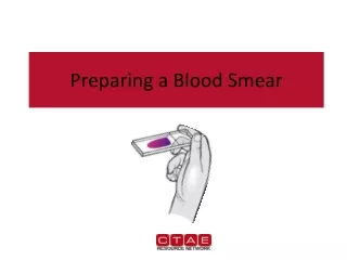
Preparation of blood smear • There are three types of blood smears: • The cover glass smear. • The wedge smear . • The spun smear. • The are two additional types of blood smear used for specific purposes • Buffy coat smear for WBCs < 1.0×10⁹/L • Thick blood smears for blood parasites .
WEDGE BLOOD SMEAR • Specimen : EDTA blood within 2 to 3 hours & collected to the mark on tube. • Not's :May change RBCs morphology such as Spiculated (crenated) cells if : • Excessive amount of anticoagulant to specimen • Old blood - long standing. • Warm environment (room temperature) may hasten changes.
Procedure • placing a drop of blood from mixed sampleon a clean glass slide. • Spreader slide using another clean glass slide at 30-40 degree angle. • Control thickness of the smear by changing the angle of spreader slide • Allow the blood film to air-dry completely before staining. (Do not blow to dry. The moisture from your breath will cause RBC artifact).
high HCT small angle low HCT large angle
CHARACTERISTICS OF A GOOD SMEAR • Thick at one end, thinning out to a smooth rounded feather edge. • Should occupy 2/3 of the total slide area. • Should not touch any edge of the slide. • Should be margin free, except for point of application.
MORPHOLOGIC CHANGES DUE TO AREA OF SMEAR • Thin area: Spherocytes which are really "spheroidocytes" or flattened red cells. True spherocytes will be found in other (Good) areas of smear. • Thick area: Rouleaux, which is normal in such areas. Confirm by examining thin areas. If true rouleaux, two-three RBC's will stick together in a "stack of coins" fashion.
Common causes of a poor blood smear • Drop of blood too large or too small. • Spreader slide pushed across the slide in a jerky manner. • Failure to keep the entire edge of the spreader slide against the slide while making the smear. • Failure to keep the spreader slide at a 30° angle with the slide. • Failure to push the spreader slide completely across the slide. • Irregular spread with ridges and long tail: Edge of spreader dirty or chipped; dusty slide • Holes in film: Slide contaminated with fat or grease • Cellular degenerative changes: delay in fixing, inadequate fixing time or methanol contaminated with water.
Biologic causes of a poor smear • Cold agglutinin: RBCs will clump together. Warm the blood at 37° C for 5 minutes, and then remake the smear. • Lipemia: holes will appear in the smear. There is nothing you can do to correct this. • Rouleaux:RBC’s will form into stacks resembling coins. There is nothing you can do to correct this
Slide fixation and staining
Romanowsky staining • Leishman's stain : a polychromatic stain • Methanol : fixes cells to slide • methylene blue stains RNA,DNA • blue-grey color • Eosin stains hemoglobin, eosin granules • orange-red color • pH value of phosphate buffer is very important
Procedure • Thin smear are air dried. • Flood the smear with stain. • Stain for 1-5 min. Experience will indicate the optimum time. • Add an equal amount of buffer solution and mix the stain by blowing an eddy in the fluid. • Leave the mixture on the slide for 10-15 min. • Wash off by running water directly to the centre of the slide to prevent a residue of precipitated stain. • Stand slide on end, and let dry in air.
CAUSES & CORRECTION • Too Acid Stain: • insufficient staining time • prolonged buffering or washing • old stain • Correction: • lengthen staining time • check stain and buffer pH • shorten buffering or wash time
Too Alkaline Stain: • thick blood smear • prolonged staining • insufficient washing • alkaline pH of stain components • Correction : • check pH • shorten stain time • prolong buffering time
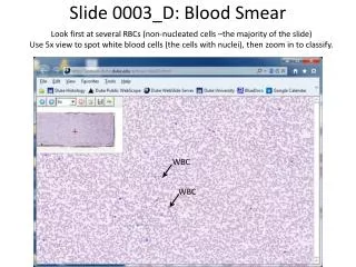
Principle • White Blood Cells • Check for even distribution and estimate the number present (also, look for any gross abnormalities present on the smear). • Perform the differential count. • Examine for morphologic abnormalities.
Red Blood Cells, Examine for: • Size and shape. • Relative hemoglobin content. • Polychromatophilia. • Inclusions. • Rouleaux formation or agglutination • Platelets. • Estimate number present. • Examine for morphologic abnormalities.
Observations Under 10X • Check to see if there are good counting areas available free of ragged edges and cell clumps. • Check the WBC distribution over the smear. • Check that the slide is properly stained. • Check for the presence of large platelets, platelet clumps, and fibrin strands.
Observing direction: Observe one field and record the number of WBC according to the different type then turn to another field in the snake-liked direction *avoid repeat or miss some cells
WBC estimationUnder 40X • Using the × 40 high dry with no oil. • Choose a portion of the peripheral smear where there is only slight overlapping of the RBCs. • Count 10 fields, take the total number of white cells and divide by 10. • To do a WBC estimate by taking the average number of white cells and multiplying by 2000.
Platelet estimationUnder 100X • Use the oil immersion lens estimate the number of platelets per field. • Look at 5-6 fields and take an average. • Multiply the average by 20,000. • Note any macroplatelets. • Platelets per oil immersion field (OIF) • <8 platelets/OIF = decreased • 8 to 20 platelets/OIF = adequate • >20 platelets/OIF = increased
Manual differential counts • These counts are done in the same area as WBC and platelet estimates with the red cells barely touching. • This takes place under × 100 (oil) using the zigzag method. • Count 100 WBCs including all cell lines from immature to mature. • Reporting results • Absolute number of cells/µl = % of cell type in differential x white cell count
If 10 or more nucleated RBC's (NRBC) are seen, correct the • White Count using this formula: • Corrected WBC Count = • WBC x 100/( NRBC + 100) • Example :If WBC = 5000 and 10 NRBCs have been counted • Then 5,000× 100/110 = 4545.50 • The corrected white count is4545.50.
Diameter:12-16 Cytoplasm : pink Granules: primary secondary Nucleus: dark purple blue dense chromatin Stab neurophile
Diameter: 12-16 Cytoplasm : pink Granules: primary secondary Nucleus: dark purple blue dense chromatin 2-5 lobes Segmented neurophile
Diameter: 14-16 Cytoplasm : full of granules Granules: large refractile, orange-red Nucleus: blue dense chromatin 2 lobes like a pair of glass Basophils
Diameter: 14-16 Cytoplasm : pink Granules: dark blue –black obscure nucleus Nucleus: blue Eosinophils
Diameter: small 7-9 large 12-16 Cytoplasm: medium blue Granules: small agranular large a few Nucleus: dark blue \round dense chromatin Lymphocyte
Diameter: 14-20 Cytoplasm : grey blue Granules: dust-like lilac color granules Nucleus: blue large irregularly shaped and folded Monocytes
LEFT-SHIFT AND RIGHT-SHIFT OF NEUTROPHIL: • Left-shift: non-segmented neutrophil > 5% (Increased bands Means acute infection, usually bacterial). • Right-shift: hypersegmented neutrophil >3%(Increased hypersegmented neutrophile )
Leukocytosis, a WBC above 10,000, is usually due to an increase in one of the five types of white blood cells Neutrophilic leukocytosis neutrophilia Lymphocytic leukocytosis lymphocytosis Eosinophilic leukocytosis eosinophilia Monocytic leukocytosis monocytosis Basophilic leukocytosis basophilia