Download
1 / 15
170 likes | 443 Views
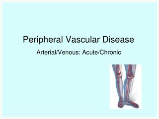
Peripheral Vascular & Lymphatic. Arteries. Carry freshly oxygenated blood Pulse – all arteries have a pressure wave / can only be felt at sites where the artery lies close to the skin and over a bone Temporal, Carotid, Brachial, Radial, Femoral, Popliteal, Posterior Tibial, Dorsalis Pedis.

E N D
Arteries • Carry freshly oxygenated blood • Pulse – all arteries have a pressure wave / can only be felt at sites where the artery lies close to the skin and over a bone • Temporal, Carotid, Brachial, Radial, Femoral, Popliteal, Posterior Tibial, Dorsalis Pedis
Ischemia • Deficient supply of oxygenated arterial blood to the tissue • Caused by obstruction of vessel • Partial blockage – may only be apparent with exercise • Necrosis – death to tissue from lack of oxygen, Irreversible
Veins • Parallel to the arteries but closer to the skin • Drain the deoxygenated blood & its waste products from the tissue & return it to the heart • No pulse, blood moves by: skeletal muscle contraction, Inspiration, Intraluminal valves
Lymphatics • Separate vessel system / retrieves excess fluid from the tissue & returns it to the bloodstream • Edema occurs without lymphatic drainage (fluid would build up)
Aids to the Lymphatic System: Spleen Tonsils Thymus gland Peyer’s Patches Bone Marrow Related Organs
The Aging Adult • Arteriosclerosis – blood vessel become more rigid causing a rise in systolic B/P • Atherosclerosis – Fatty plaque accumulation in the intima of arteries • Progressive enlargement of calf veins w/prolonged inactivity & heart failure leads to risk of DVT & Pulm. embolism
Inspect & Palpate • Leg pain or cramps • Claudication (Limping) Distance - # of blocks or stairs that produces pain • Temp – coolness is assoc. w/ arterial disease • Edema – bilateral w/ CHF (systemic) unilateral w/local obstruction or inflammation
Palpate • Homan’s Sign – calf pain with dorsiflexion (may be due to DVT) • Palpate & Grade the Pulses • Pitting Edema – Bilateral occurs w/ CHF, Diabetic neuropathy, Hepatic cirrhosis (scale of 1+ to 4+)
Raynaud’s Syndrome • Bilateral response to cold, vibration, or stress • 1st white – arteriospasm • 2nd blue – cyanosis as spasm relaxes • 3rd red – return of blood to capillaries
Lymphedema • Damage or removal of lymph nodes impedes drainage of lymph • Unilateral swelling, non pitting
Deep Vein Thrombosis (DVT) • Deep vein occluded by:thrombus/clot • Sudden onset, pain w/dorsiflexion • Positive Homan’s sign – accurate < ⅓ of the time • Risk for PE (Pulmonary Embolism)
Aneurysm • Usually caused by atherosclerosis which weakens lining of vessel • Aorta – most common site • Effects of Blood Pressure causes ballooning of the vessel • More common in males and men over 55 & women over 70
Inspect & Palpate • Clubbing , • Capillary Refill – reflects peripheral perfusion & cardiac output <2 sec. • Upper arm edema – lymph obstruction • Grade & Compare Pulses, 2 + is normal • Homan’s Sign – calf pain w/
General condition Arterial assessment * BP both arms, palpate pulses for amplitude Auscultate for bruit over carotid Inspect & palpate extremities for arterial versus venous problems Estimate the jugular venous pressure Check for Homan’s sign & edema PERIPHERAL VASCULAR SYSTEM