Download
1 / 32
340 likes | 632 Views
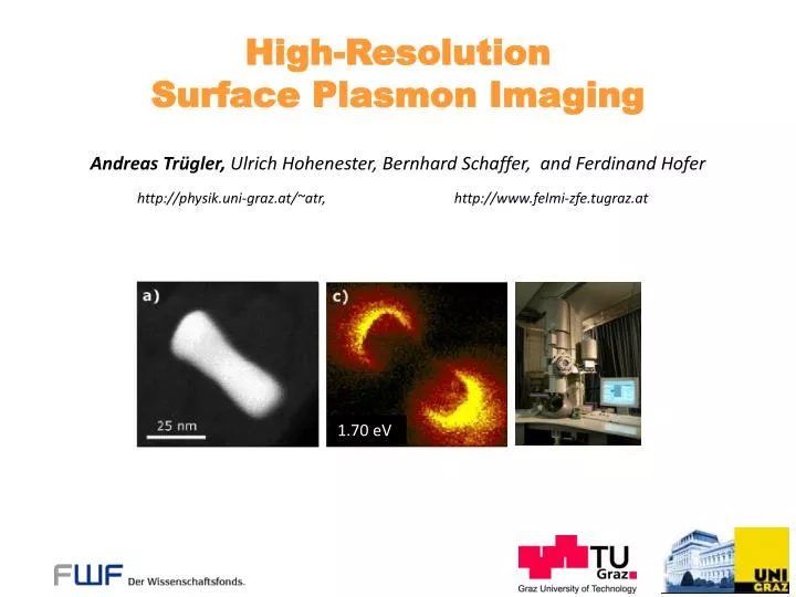
1.70 eV. High-Resolution Surface Plasmon Imaging. Andreas Trügler, Ulrich Hohenester, Bernhard Schaffer, and Ferdinand Hofer. http://physik.uni-graz.at/~atr, http://www.felmi-zfe.tugraz.at.

E N D
1.70 eV High-Resolution Surface Plasmon Imaging Andreas Trügler, Ulrich Hohenester, Bernhard Schaffer, and Ferdinand Hofer http://physik.uni-graz.at/~atr, http://www.felmi-zfe.tugraz.at
W. Barnes et al., Surface plasmon subwavelength optics, Nature 424, 824 (2003). H. Atwater, The promise of plasmonics, Scientific American 296(4), 56 (2007). „Flat light“ Particle plasmons Meta materials Agenda: Theoretical background and how to map a plasmon Simulation technique Comparison between experiment and theory The promise of plasmonics...
Theoretical background and how to map a plasmon
No! Because the wavelength of light is (much) larger than the nanoparticle but: What about other microscopy techniques? EELS Electron Energy Loss Spectroscopy EFTEM Energy Filtered Transmission Electron Microscopy F. J. García de Abajo : Optical excitations in electron microscopy, arXiv:0903.1669v1 (2009) Can one „directly see“ surface plasmons?
Electron beam excites surface plasmon (evanescent source of radiation) Surface plasmon acts back on electron ~ 100 keV electrons ( approx. 70 % of c ) Raster scanning of electron beam probes dielectric environment of MNP Electron interaction
Maxwell‘s equations: Solution by Green’s functions: Structure of equations we want to solve: Green function equation: 4-vector notation, . Liénard-Wiechert potentials
Maxwell‘s equations: Solution by Green’s functions: Structure of equations we want to solve: Solution for an arbitrary charge distribution: 4-vector notation, . Liénard-Wiechert potentials
Solution for a relativistic moving charge: Charge distribution of an electron moving along r(t): Liénard-Wiechert potentials
Solution for a relativistic moving charge: Charge distribution of an electron moving along r(t): Liénard-Wiechert potentials
Solution for a relativistic moving charge: Charge distribution of an electron moving along r(t): Liénard-Wiechert potentials
Solution for a relativistic moving charge: Charge distribution of an electron moving along r(t): Liénard-Wiechert potentials
Solution for a relativistic moving charge: Charge distribution of an electron moving along r(t): Final potentials: Liénard-Wiechert potentials
Energy loss of the fast electron: Energy loss can be related to the work against the induced electric field! Fourier transform Process reminiscent of a self energy. Probe of the electrost. potential of the SP! U. Hohenester, H. Ditlbacher, and J. R. Krenn: On the interpretation of electron energy loss spectra of plasmonic NPs (to be published) ~ 100 keV electrons Electron energy loss
Energy loss of the fast electron: Energy loss can be related to the work against the induced electric field! Fourier transform Loss probability: The problem reduces to solving the electric field induced by the electron: Electron energy loss
Fourier transform: Time domain: Electron beam interacts with a SP oscillating in time. Frequency domain: The SP oscillation becomes frozen, and interacts with a periodically modulated charge distribution of the electron beam. (Interaction along the whole trajectory!) Change of the reference frame
Electric Green tensor: The electromagnetic response of a structured material is fully captured in its electric Green tensor. Green tensor - link to simulation
Mie theory Analytic results for spherical particles to test the simulation. Boundary Element Method (BEM) Approximate surface of scatterer by small surface elements. Works for scatterers which have a homogeneous dielectric function. Up to a few 1000 surface elements. Simulation of particle plasmons
1. Discretizationof particlesurface Using standard triangulation techniques of Matlab®with typically a few thousand surface elements Simulation of particle plasmons
2. Excitationof nanoparticle ( illumination, molecule, electron beam ... ) Oscillating dipole emetal(w), eb Inside and outside the metallic nanoparticle Maxwell‘s equation are the usual wave equations!The only non – trivial contribution comes from the boundaries. Simulation of particle plasmons
3. Add surface charges and currents such that BC of Maxwell‘s equations are fulfilled Oscillating dipole Boundary Element Method approach (BEM ) Garcia de Abajo & Howie, PRB 65, 115418 (2002); Hohenester & Krenn, PRB 72, 195429 (2005). Simulation of particle plasmons
Results and comparison between experiment and theory
Inelastically scattered electrons EELS, EFTEM Incident high energy electrons 60-300 kV Auger electrons X-rays Secondary electrons Thin specimen 10-200 nm Elastically scattered electrons TEM, HREM, ED Ferdinand Hofer and Bernhard Schaffer, Austrian Centre for Electron Microscopy and Nanoanalysis (FELMI) , TU Graz Transmission electron microscopy
Gold particles (low-loss) ZLP – zero loss peak (energy resolution: FWHM) plasmon peaks HAADF ZLP: Measures energy distribution of primary electron beam, defines resolution Monochromated STEM-EELS
Experiment Theory J. Nelayah, M. Kociak , O. Stéphan, F. J. García de Abajo, M. Tencé, L. Henrard , D. Taverna, I. Pastoriza-Santos, L. M. Liz-Marzán, and C. Colliex, Mapping surface plasmons on a single metallic nanoparticle, Nature Phys. 3, 348 (2007). Surface plasmon mapping with EELS
1.0eV 1.6eV 2.4eV Image size: Energy range: Slit width: Energy steps: Energy resolution: Acq. time: 512 x 512 px -1 to 4.5 eV 0.3 eV 0.1 eV ~400 meV 17 min Narrow slit (0.3 eV) combined with monochromator gives an energy resolution of ~0.4 eV. This allows EFTEM imaging close to the zero-loss peak, showing SP modes of Au nanoparticles. Schaffer et al., Micron (2008), DOI:10.1016/j.micron.2008.07.004 Low-loss mapping by EFTEM
64 x 64 px -2 to 8 eV 220 meV 54 min Image size: Energy range: Energy Res.: Acq. time: Image size: Energy range: Energy Res.: Acq. time: 512 x 512 px -1 to 4 eV 450 meV 15 min @1.8 eV @ 1.8 eV @1.0 eV @ 1.0 eV Monochromated EFTEM Monochromated STEM EELS EELS and EFTEM results
Comparison with Simulation 1 2 3 @ 1.08 eV 400 nm @ 1.85 eV @ 2.29 eV 75 nm B. Schaffer, U. Hohenester, A. Trügler, and F. Hofer Phys. Rev. B 79, 041401(R) (2009) EELS and EFTEM results
Comparison with Simulation 1.08 eV 1.85 eV 2.29 eV (0.80 eV) (1.50 eV) (2.33 eV) 1 2 3 @ 1.08 eV 400 nm @ 1.85 eV @ 2.29 eV 75 nm B. Schaffer, U. Hohenester, A. Trügler, and F. Hofer Phys. Rev. B 79, 041401(R) (2009) EELS and EFTEM results
Comparison with Simulation 1.08 eV 1.85 eV 2.29 eV (0.80 eV) (1.50 eV) (2.33 eV) EELS and EFTEM results
Theoretical description Liénard-Wiechert potentials Electromagnetic Green tensor link to simulation Simulation Discretize particle surface boundary element method Results Very nice agreement with EELS & EFTEM measurements Direct observation of surface plasmons with unmatched spatial resolution! Summary
Theoretical Nanoscience (KFU) Ulrich Hohenester Hajreta Softic Jürgen Waxenegger Nanooptics (KFU) Thank you for your attention! Joachim Krenn Alfred Leitner Harald Ditlbacher Daniel Koller Andreas Hohenau Franz Aussenegg FELMI (TU Graz) Ferdinand Hofer Bernhard Schaffer