Download

1 / 56
650 likes | 1.36k Views
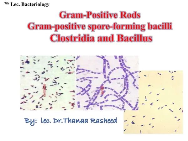
Gram Positive Rods. Listeria. Small Gram positive rods or coccobacilli (<2 μ m ) Tolerate wide temperature and pH range Small haemolytic colonies on blood agar ( β haemolysis ) Facultative anaerobes Catalase positive, oxidase negative Tumbling motility at 25 degrees Aesculin hydrolysed

E N D
Listeria • Small Gram positive rods or coccobacilli (<2μm) • Tolerate wide temperature and pH range • Small haemolytic colonies on blood agar (β haemolysis) • Facultative anaerobes • Catalase positive, oxidase negative • Tumbling motility at 25 degrees • Aesculin hydrolysed • Do not grow on MacConkey agar • Environmental • Outbreaks related to silage
Listeria spp L. monocytogenes meningoencephalitis septicaemia abortion pyogenic infection L. ivanovii abortion, systemic infection L. innocua non-pathogenic L. seeligeri “ L. welshimeri “ L. grayi “
Epidemiology • Common commensal (tonsils, intestine) and environmental organisms • grow at temperatures 4 - 45°C, pH 6.0 - 9.6 • Incidence relates to management/husbandry, (silage feeding), seasonal • predisposed by trauma, immunocompromise, hormonal alterations
Diagnosis • Specimens: • Visceral form: liver, kidney or spleen • Neural form: spinal fluid, brain stem • Abortion: placenta, foetal abomasal contents • Blood smear is not very informative • Isolation: grow aerobically on both blood and MacConkey agars at 37°C for 24-48h • Colonies appear transparent after 24 hours and greyish in 48 hours. Pathogenic strains show β haemolysis • L. Monocytogenes is CAMP +ve with Staph aureus • L. Ivanovii is CAMP +ve with Rhodococcus equi CAMP test
Listeriosis - Ruminants • Meningoencephalitis • Most common form = circling disease (small ruminants) • animal circles in one direction only • unilateral facial paralysis, difficulty in swallowing • fever, blindness, headpressing • paralysis, death in 2 - 3 days
Listeriosis - Ruminants • In pregnant animals, may localize in placentomes • cross-over to amniotic fluid, multiplies • ingested by fetus, causes fetal death, abortion • In milking cows, mammary gland can be involved • subclinical mastitis, contamination of milk • may survive low temp pasteurization inside MØ • lengthy survival in nature • growth at low temperatures • Entry also by nasal mucosa, conjunctivae • Direct access to nervous system via dental plates of trigeminal ganglia
Pathogenesis • tooth loss & cutting naïve/neonatal pregnancy animal • • oral inoculation epithelial invasion • • trigeminal nerve bacteraemia • • brain stem neonatal placentitis • septicaemia • meningoencephalitis abortion • “circling disease”
Silage and sheep Listeria encephalitis (bacteria in brain) A –Circling disease in sheep B – Cranial nerve paralysis
Pathogenic mechanisms • Facultative intracellular parasites surviving in macrophages and epithelial cells • Cell uptake induced by bacterial protein internalin (inlA) • Inside the cell they escape the phagolysosome, multiply in the cytoplasm and via actin based motility spread laterally to adjacent cells • They escape epithelium and are taken up by PMN and macrophages • These cells are killed and the organism may spread systemically
Pathogenic mechanisms • Bacteria polymerize actin, form tails • hollow mesh forms on surface, left behind as bacterium moves through cytoplasm, invade adjacent cells • actin depolymerized as organism moves (turnover) • Major virulence factor mediating intracellular survival is a cholesterol binding cytolysin called listeriolysin (LLO) • Shares 40-50% aa similarity to other thiol activated toxins • Mediates escape from phagocytic vesicle
LLO • Cholesterol-directed pore-forming cytolysin (>22 members) • Bind to cholesterol-containing membranes • Insertion • Oligomerization (20-80 mers) • Pore (20-30 nM) formation
Epidemiology of Human Listeriosis • enteric Listeria in animals • • contamination of carcase, milk or food crops • • Ingestion (pate, soft cheese, coleslaw) • • colonisation of tonsils and intestine • • immunocompetent immunocompromised • (neonate, elderly, pregnancy) • • asymptomatic septicaemia • meningitis • abortion
Associated Foods • Milk products – raw pasteurised • Cheeses • Meat and poultry products – Raw, cooked ready to eat meat and poultry (sporadic and epidemic) • Seafood – fresh, frozen and processed seafood
Consequences of human L. monocytogenes infection • Entry via GI tract, ~ 20 h incubation period • usually asymptomatic/mild, influenza-like symptoms in adult humans, transient gastroenteritis • more serious infection immunocompromised • CNS infections (encephalitis, meningitis), fatal bacteremia • puerperal sepsis • crosses placenta in utero fetal infection • stillbirths, preterm labor • infant born with systemic infection
Cutaneous listeriosis (in vets) • 17 cases, all with lesions on fore or upper arms, or hands • 16 farmers, veterinarians • 1 butcher • Most developed lesions 1-4 days after attending congenitally infected bovines
Erysipelothrix rhusiopathiae • Gram positive rods (1-2.5μm) • Commensal: widespread in animals, infects man • Grows: 4-37°C • Catalase negative, oxidase negative • Facultative anaerobic, non-motile • Opaque, pin-point, non-haemolytic colonies • infection & disease: mainly in pigs; sheep, turkeys, others • Smooth (S) and rough (R) forms associated with different diseases • Acute septicaemia in pigs, turkeys, sub-acute skin lesions in pigs – S • Chronic arthritis in sheep, endocarditis in pigs - R
Pathogenesis commensal: tonsils, RES, bone marrow, many organs mainly R or R/S depression of host defences multiplication of virulent strains (reversion to S?) entry via tonsils or cuts/abrasions invades neutrophils septicaemia urticarial form arthritis/endocarditis fever malaise persistent anorexia fever erosive DIC DIC chronic inflammation haemorrhage lymphadenitis epidermal lesions fatal ("Diamonds")
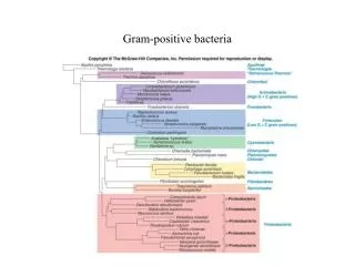
Actinomycete Group • Gram positive • Catalase positive • Many are acid fast (mycolic acid in cw) • Slow growing - survive in macrophages • Pleomorphic (coccobacilli to filamentous). Very small (<1μm) • Many saprophytic – some opportunist • Pathogenic genera Actinomyces spp. Nocardia spp. Arcanobacterium spp. Dermatophilus spp. Corynebacterium spp. Eubacterium spp. Rhodococcus spp.
Actinomycete diseases • Pyogenic • Granulomatous • Include: abscesses, pyelonephritis, lymphadenitis, osteomyelitis • Chronic inflammation – focal lesions • Some highly-host adapted • CMI protective – A/bodies mainly non-protective • In vitro - sensitive to some antibacterials • In vivo – poor response - intracellular location
Actinomyces • Actinomyces bovis, Actinomyces viscosus, Actinomyces suis • (Most) non-acid fast branching rods • non-motile • microaerophilic or anaerobic • Colonies are non-haemolytic, small and white • Produce pyogenic, granulomatous reactions - “sulphur granules”
Actinomyces bovis • Normal flora - anaerobic • Thick yellow pus – “sulphur granules” • (occ. confused with wooden tongue) • Causes actinomycosis or Lumpy jaw in cattle (other tissues). Invasion through wound/rough feed/damaged mucosa – Bone infection – osteomyelitis – animal stops eating, loses weight • Soft tissue infection • Mastitis
Actinomyces bovis • Infection endogenous • Organism lives in the mouth normally • Pathology the result of tissue trauma, lesions or prolonged irritation • Treat – lesions small, circumscribed – surgery, abscesses drained, packed with gauze and iodine + penicillin
Gram stain,A. bovis Sulphur granules 48 hour culture, A. bovis
Actinomyces viscosus • Mainly dogs (cats, pigs, goats, cattle, horses). • Fimbriae - adherence to teeth – plaque • Similar lesions to Nocardia (Nocardia rarely produces granules) • Localised, pyogranulomatous lesions • Two main conditions - Thoracic lesions (pleural, pericardial fluid, lung lesions) and osteomyelitis • Treatment – prolonged Actinomyces suis – mastitis in pigs (sows), trauma initiates disease
Nocardia • Strictly aerobic • Gram-positive, pleomorphic (filaments, rods, cocci – branching) • Widely distributed – soil, water, air, sewage • 0.5 – 1.2 μm in size • Acid fast • Non-motile • Non-spore-forming
Nocardia • 12 species – pathogenic to birds, goats, cats, dogs, fish, horses, cattle and humans • Nocardia asteroides most frequent nocardial pathogen – subcutaneous infections in dogs • N. brasiliensis – pneumonia in horses • Colonies are vivid white – occasionally pigmented
Nocardia • Organism inhaled, in wound or ingested • Direct or haematogenous spread • Prevents phagocytosis • Chronic invasive pyogenic infections (no sulphur granules) • 3 clinical forms – cutaneous, respiratory (pyothorax) and systemic (pyrexia, cough, neurological, resembles distemper) • Dogs – 3x more common in males, Cats – mainly thoracic infection • Treatment: difficult and prolonged – not penicillin
Dog, Nocardia Fluid from chest 4 day culture from fluid
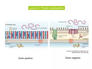
Dermatophilus congolensis • Facultatively anaerobic • Gram positive, non acid fast • filamentous, branching – filaments mature they fragment and release motile flagellate spores • Septation of filaments – zoospores (motile) Beaded, ‘Tram-track’ – the infectious form • CO2chemotactic to zoospores – zoospores germinate form filament form new zoospores – repeating the cycle Pathogenesis • Commensal/spore entry – injury/ wet damage • Colonisation – keratinase production. Aid spread and growth • Strong host response, neutrophil and lymphocyte exudation. • EPIDERMIS ONLY
Dermatophilus congolensis • Dermatitis – cattle, dog, cat, man • Sheep – ‘lumpy wool’, ‘strawberry foot’ • Horse – ‘mud fever’, ‘greasy heel’, ‘rain scald’ • Treatment – Pen/Strep. Tetracyclines • No effective vaccine Dermatophilus in a horse Dermatophilus lesions, SHEEP
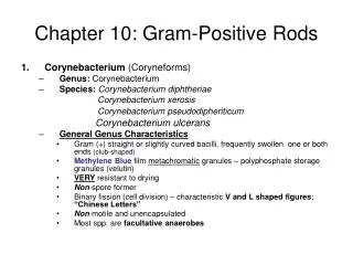
Corynebacterium • Small pleomorphic, Gram-positive rods • Chinese letters • Facultatively anaerobic, non-motile • Catalase positive, oxidase negative • Pyogenic • Common commensals • Colonies are white, small, dry, non-haemolytic (except C.pseudotuberculosis). • Genus originally created for the important human pathogen C. diphtheriae, the cause of diphtheria in man
CAMP Test CAMP-Inhibition Test 1. Staphylococcus aureus 2. C.pseudotuberculosis 3. C.renale 4. Rhodococcus equi
Corynebacterial species of veterinary interest C. renale group: • C. renale • C. pilosum • C. cystitidis All cause cystitis and pyelonephritis in cattle Diphtheria group: • C. diphtheriae • C. ulcerans • C. pseudotuberculosis Various diseases of cats, cattle, horses, small ruminants and humans
Corynebacterium renale group • C. renale > C. cystitidis > C. pilosum • Opportunist – highly adapted • Causes cystitis, pyelonephritis, balanoposthitis • Predisposing factors - pregnancy, parturition, post mating • 90% bulls – C. cystitidis - prepuce
Cow with C. renale infection Urine, C. renale C. renale on milk agar
Corynebacterium renale group • Pathogenesis • Adhere to urogenital mucosa • “Stress” • Proliferation • Ascending infection • Inflammation • Cystitis/pyelonephritis • Virulence factors Pili - adherence Renalin - cell lysis Urease Caseinase
The “diphtheria group” • Highly-related (based on DNA hybridisation studies) • C. diphtheriae – diphtheria (humans) • C. pseudotuberculosis – various pyogenic infections • C. ulcerans – nasal congestion (cats), mastitis (cattle) • Susceptible to infection with β-corynephages • C. pseudotuberculosis +C. ulcerans zoonotic pathogens • C. diphtheriae normally only a human pathogen
Corynebacterium diphtheriae • Occasionally isolated from infected wounds in horses (potential environmental reservoir?) • Ironically, horses were the original heroes in fight against diphtheria An illustration from the Nov. 17, 1894, issue of Scientific American, showing doctors drawing blood from a horse to produce antitoxin for diphtheria
Corynebacterium ulcerans • Significant increase in human infections caused by C. ulcerans • Same organism isolated from several domestic cats with bilateral nasal discharge (2002-2003) • Strains isolated from domestic cats were found to exhibit the predominant types observed among human clinical isolates, suggesting that C. ulcerans isolated from cats could be a potential reservoir for human infection
Corynebacterium pseudotuberculosis • C. pseudotuberculosis (pseudes-tuberculosis) • Thought to have spread from Europe with expanding colonial powers • 2 biotypes identified: • “ovis”: non-nitrate reducing, infect sheep/goats (CLA) • “equi”: nitrate reducing, infects predominantly horses