Download
1 / 22
220 likes | 430 Views
Cardiovascular System: Blood Vessels and Circulation. A Presentation. Structure and Function. The vital functions of the cardiovascular system occur at the capillary level Chemical and gaseous exchange between the blood and interstitial fluid occurs across capillary walls.

E N D
Cardiovascular System: Blood Vessels and Circulation A Presentation
Structure and Function • The vital functions of the cardiovascular system occur at the capillary level • Chemical and gaseous exchange between the blood and interstitial fluid occurs across capillary walls. • Tissues relay on capillary diffusion for nutrients and oxygen and to remove metabolic waste.
Structure of vessel walls • Three layers • Tunica intima or innermost layer. Includes a lining of endothelium and a connective tissue layer. • Tunica media or middle layer. Contains smooth muscle tissue and collagen and elastic fibers. • Tunica externaforms sheath of connective tissue around the vessel.
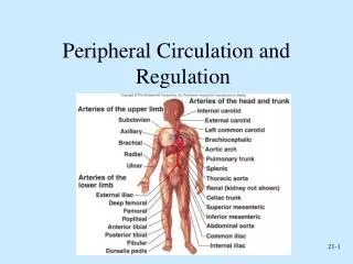
Arteries • Elastic: large, able to absorb the pressure changes of the cardiac cycle, contain many elastic fibers that stretch and return to original dimension. Examples: aorta, pulmonary trunk • Muscular arteries: medium sized distribute blood to skeletal muscles and internal organs. Not as elastic. Example: external carotid arteries.
Arterioles: small (30u), diameter of muscle layer very thin.
Capillaries • Only vessels that permit exchange between blood and interstitial fluid. • Walls are thin(no tunica externa or tunica media) • Small diameter slows blood flow • Permits water, small solutes and lipid-soluble materials to pass. • Interconnect to form capillary bed.
Veins collect blood and return it to the heart • Venules: smallest • Veins have relatively thin walls. • In medium veins contain valves that prevent the backflow of blood due to low pressure and gravity • Stretching and distortion of these valves cause varicose veins.
Pressure and resistance determine blood flow and affect rates of capillary exchange • Highest pressure at the base of the aorta • Resistance opposes movement of blood. • Sources include • Vascular resistance • Viscosity • turbulence
Pressures within the systemic circuit • Highest in the aorta and lowest at the vena cava • Arterial pressure rises during ventricular systole and falls during ventricular diastole. • 120/80 reflects the separate systolic and diastolic pressures. • Read the Clinical Note page 435.
Venous pressure • Pressure is only 1/10th of the arterial system at the beginning. • When standing two factors help venous return • Muscular compression of skeletal muscles • Respiratory pump: inhalation causes both the vena cava and rt. atria to expand and fill.
Cardiovascular Regulation • Involves • Autoregulation • Neural mechanisms • Endocrine mechanisms
Pulmonary Circuit • Deoxgenated blood enters the lungs in arteries • Oxygenated blood leaves the lungs in veins.
Systemic Circuit • Oxygenated blood from the left ventricle goes to tissues other than the lungs’ exchange surfaces. • Deoxygenated blood returns to the right atrium.
Fetal Circulation • Embryonic lungs are collapsed and nonfunctional • All nutritional and respiratory needs are provided by diffusion across the placenta. • Umbilical arteries carry deoxygenated blood from fetus to placenta • Umbilical veins returns oxygenated blood from placenta to fetus.
Veins bypass developing liver through ductusvenosus. • Foramen ovalein fetal heart and ductusarteriosus between pulmonary and aortic trunks by pass collasped lungs. • At birth lungs expand and smooth muscles in ductusarteriosus contract closing connection. Increased pressure in L atrium closes foramen ovale.
Aging • Decreased hematocrit • Blockage of peripheral veins • Pooling of blood in the veins • Reduction in max. cardiac output • Changes in heart conduction • Reduction in elasticity of cardiac skeleton • Progressive atheroschlerosis • Replacement of damaged heart muscle by scar tissue.
Template Provided By www.animationfactory.com 500,000 Downloadable PowerPoint Templates, Animated Clip Art, Backgrounds and Videos