Download

1 / 41
410 likes | 639 Views
Burns. Nicole Baier, MD. Statistics. In US: 1.2 million burns each year 60,000 hospitalizations 6000 deaths 2 nd leading cause of unintentional death in children (after MVA) Pediatric incidence by type of burn: Scald burns: 85% Flame burns: 13%

E N D
Burns Nicole Baier, MD
Statistics • In US: • 1.2 million burns each year • 60,000 hospitalizations • 6000 deaths • 2nd leading cause of unintentional death in children (after MVA) • Pediatric incidence by type of burn: • Scald burns: 85% • Flame burns: 13% • Remaining 2%: electrical and chemical burns
The Skin epidermis dermis • Barrier to: • Fluid loss • Entry of infection • Heat loss
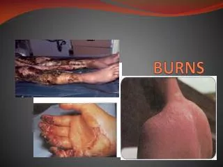
Burn Classification • 1st degree (superficial) • Epidermis only • Erythematous, painful • No blistering
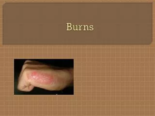
Classification • 2nd degree (partial thickness) • Injury to epidermis and variable portion of dermis • Moist, pink or red, blanches to touch • Vesicles and blisters • Extremely painful • Heal spontaneously
Classification • 3rd degree (full thickness) • Entire epidermis and dermis • No residual epidermal cells – require skin grafting • Leathery, white or black or brown • Not painful (no viable nerve endings) • High risk of scarring
Classification • 4th degree • Involve underlying structures (tendons, nerves, muscles, bone, fascia) • Reconstructive surgery often necessary
Estimation of Burn Size • Used to calculate fluids for IVF resuscitation • Only 2nd and 3rd degree burns considered • Adults: Rule of nines • Pediatric: Lund-Brower chart • Estimate: palm of patient’s hand = 1% BSA
Acute Assessment • AIRWAY • Airway edema caused by inhalational injury • Direct thermal injury – supraglottic • Suspicion increased if: • Facial/ oral burns • Soot in mouth/nose • Singed nasal hairs • Wheezing, stridor, or hoarseness noted • Intubation should be performed quickly as edema can progress rapidly (over initial 24-36 hours)
Acute Assessment • BREATHING– Initial findings • Early hypoxia may result from: • Airway obstruction • Impaired chest wall compliance (circumferential burns) • Decreased ambient FiO2 (10-15%) • Carbon monoxide • Cyanide • Produced when wool, silk, nylon, polyurethane burn • Disrupts mitochondrial oxygen use by complexing with cytochrome • CO and CN are responsible for majority of early mortality at scene • Children more susceptible to toxicity of inhaled materials due to higher minute ventilation
Carbon Monoxide • Affinity for hemoglobin 250x > O2 • Decreases oxygen carrying capacity • Shifts oxyhemoglobin dissociation curve to left • Binds to myoglobin and mitochondrial cytochrome oxidase • Interfere with cell oxygen use and energy production • Measured with co-oximetry • 20-30% = headache, dizziness • 40-50% = altered LOC • >50% = coma, death • Treatment: 100% oxygen • ½ life in room air: 4-6 hours • ½ life in 100% FiO2: 40-60 minutes
Acute Assessment • BREATHING – Later findings • Chemical irritants injure tracheobronchial tree and lung parenchyma • Lower airway edema • Respiratory epithelium sloughs - cast formation causes airway obstruction • Manifests as: bronchospasm, post-obstructive atelectsis • Patients also at risk for: • Surfactant deficiency due to damage to type II pneumocytes • ARDS • After 72 hours: nosocomial pneumonia may develop • Restrictive lung disease may develop in survivors
Acute Assessment • CIRCULATION • In 50% BSA burn: • 1 minute after burn, cardiac output is ½ of preburn state • At 1 hour, cardiac output is 1/3 of preburn state • Hypovolemic shock • Loss of skin integrity increases evaporative losses 6-7X • Increased vascular permeability leads to interstitial edema and intravascular volume loss • Maximal at 30 minutes • Capillary integrity restored 8-12 hours post-injury • Myocardial depression also occurs • Thought to be due to TNF release
Acute Management • CIRCULATION • Burns >15% BSA require IV fluid resuscitation to maintain perfusion • Time to IV access is a major predictor of mortality in pediatric patients who have burns greater than 80% TBSA • IV preferably placed in nonburned tissues
Acute Management • CIRCULATION • Parkland Formula: • Used to determine resuscitation fluids = LR • 4 mL x weight (kg) x % TBSA burned • ½ over 1st 8 hours, ½ over remaining 16 hours • Added to maintenance dextrose-containing fluids • Monitor hemodynamics, urine output and adjust fluids accordingly
Question You have a 14 month old, 11 kg infant who was involved in a house fire and has second degree burns to both of her hands, feet, her right lower arm and both lower legs. What IV fluids should she receive over the 1st 24 hours?
Answer = 26% TBSA Burn • Calculate % BSA: • Both hands: 3 x 2 = 6% • Both feet: 3.5 x 2 = 7% • Right lower arm = 3% • Both lower legs: 5 x 2 = 10% • Parkland Formula: • 4 mL x 11 kg x 26% = 1144 mL fluid resuscitation requirement • 572 mL over 1st 8 hours = 61 mL/hr of LR • 572 over remaining 16 hours = 35 mL/hr of LR • Maintenance Fluid Requirement • 44 mL/hr of D5 ½ NS
Other initial management • Remove all clothing that is hot/ burned/ exposed to chemicals • Prevent continued skin damage • Wound treatment • Clean with mild soap and water • Apply cool saline-soaked gauze – decreases pain • Do not apply ice – produces hypothermia, worsens damage • Covering with a sheet may decrease pain by decreasing environmental exposure
Electrical injuries • Minor surface burns may hide massive coagulation necrosis of muscle and deep tissues • Risk of rhabdomyolysis • Risk of cardiac abnormalities • Asystole, ventricular tachycardia/ fibrillation • Atrial and ventricular ectopy, 1st and 2nd degree heart block, bundle branch blook, prolonged QT • Non-specific ST-T changes and interval delays most common
Electrical Injuries • Tissue injury is directly proportional to resistance • Nerves, muscles, blood vessels have lowest resistance • Electricity preferentially flows through these structures • More severe damage • Increased resistance: • Skin • Tendons • Bone • Fat • Water decreases resistance, therefore moist areas (eg, axillae) tend to sustain more damage
Electrical Injuries • Type of current • AC (household electricity) is more dangerous • Continual muscle contraction and relaxation results in muscle tetany • Eg, a 60 Hz alternating current changes direction 120x/ second • DC (lightning strikes) produces muscle contraction only at beginning and end of current flow
Electrical Injuries • Current Pathway • Current may flow in 1 of 3 pathways: • Hand to hand • 60% mortality rate due to: • Spinal cord transection at C4-C8 • Suffocation due to chest wall muscle tetany • Myocardial muscle damage • Hand to foot • 20% mortality rate due to cardiac arrhythmias • Foot to foot • 5% mortality rate
Additional Management for Electrical Injuries Obtain EKG Consider obtaining cardiac enzymes Monitor patients with medium and high-voltage injuries on monitor for 24-72 hours
Compartment Syndrome • Most common early cause of diminished pulses is inadequate resuscitation • High index of suspicion for elevated compartmental pressures in circumferential burn • Emergent escharotomy or fasciotomy is indicated for limb salvage in pulseless extremity • Thoracic escharotomies are occasionally required to improve chest-wall compliance and facilitate ventilation
Ongoing Management • Hypermetabolic state • Increase in metabolism over 1st 5 days – then plateau through remainder of acute admission and into rehab • Due to surge of catecholamines, cortisol, aldosterone, growth hormone • Insulin secretion decreased, tissues insulin resistant • Degree correlates with extent of injury
Hypermetabolic State • Manifestations • Tachycardia, increased cardiac output • Hyperthermia • Baseline temp reset to 38.5⁰C • Increased gluconeogenesis, protein catabolism, lipolysis • Resting energy expenditure 2-3 x normal • May be associated with: • Impaired wound healing • Sepsis • Loss of lean body and muscle mass
Hypermetabolic State Hart DW, Wolf SE, Mlcak R, et al. Persistence of muscle catabolism after severe burn. Surgery 2000; 128: 312–319. • In burn injuries > 40% TBSA: • Resting metabolic rate at 33°C is: • 180% of basal rate at admission • 150% at full healing of the wound • 140% 6 months after the injury • 120% at 9 months post injury • 110% after 12 months
Hypermetabolic state Rutan FL, Herndon DN. Growth delay in postburn pediatric patients. Arch Surg 1990; 125: 392-395. • Long-term consequences • Profound muscle wasting • Decreased bone mineral density • Retarded linear growth in children • In 80 patients with > 40% TBSA burn: • Profound growth arrest notedduring postburn year 1 • Growth improved to normalby postburn year 3
Ongoing Management • Feeds started EARLY • Within 6 hours of admission • Require up to 50% more calories than at baseline • Hypermetabolic state • Pain and anxiety increase physiologic demands • Greater heat loss occurs in young infants with larger surface area-to-mass ratios • Reduces bacterial translocation and sepsis • TPN avoided due to infectious complications • Goal: full feeds by 24-48 hours
Infectious Concerns • Risk of infection related to: • Loss of skin barrier • Wound colonization is universal by 1-2 weeks post-injury • Presence of inhalational injury - compromises normal clearance mechanisms • 5x higher rate of pneumonia • Immunosuppression • Impaired cellular and humoral immune response • Infection now responsible for 50-60% of deaths in burn patients
Topical Therapies • Bactroban • Used for superficial burns, primarily on face • Silvadene (silver sulfadiazene) • Bacteriocidal • Cannot be used in those with sulfa allergies • Causes neutropenia and thrombocytopenia
Topical Therapies • Sulfamylon (mafenide acetate) • Better penetration of deep burns, eschars, and cartilage • Bacteriostatic • Better gram negative coverage (pseudomonas) • Causes fungal overgrowth • Painful • Carbonic anhydrase inhibitor – causes metabolic acidosis
Surgical Wound Management Hart DW, Wolf SE and Chinkes D, et al. Determinants of skeletal muscle catabolism. Ann Surg 2000; 233: 455–465. • Early excision and closure of full thickness burn wound • If wound >50% TBSA is totally excised and covered with autograft within 2–3 days: • Metabolic rate 40% less compared with wound coverage 1 week post injury
Surgical Wound Management • Other benefits of early wound excision • Decreases pain • Provides barrier to fluid and heat loss, bacterial invasion • Decreases length of stay • Accelerates recovery • Fewer septic complications • Decreased morbidity and death
Surgical Wound Management • Serial wound excision and grafting is the standard of care for full-thickness burns • When the burned area exceeds donor site supply (burns >30% BSA), homografts from donors or skin substitutes are used • Taken back to OR weekly to replace homografts with autografts as donor sites heal
Criteria for Admission • >15% BSA • 3rd degree burns • Electrical burns • Inhalational injury • Burns to hands, feet, face, genitalia, joint surfaces • Suspected abuse or neglect • Inadequate home situation
Outpatient Treatment • Leave blisters intact • Dress burns with silvadene • Wash wound and change dressings BID • Pain control with tylenol or tylenol with codeine
Identifying abusive burns • 15-20% of burn injuries are the result of abuse • Suspicious patterns: • Glove or stocking burns of hands and feet • Deep burns on trunk or back • Small-area full-thickness burns (cigarette) • Circumferential burns • Burns localized to the perineum or buttocks • Symmetric burns
Burn Prevention • Preset water heaters to max of 120⁰ F • Duration of exposure required to produce full-thickness burn: • 120⁰ F: 10 minutes • 130⁰ F: 30 seconds • 140⁰ F: 5 seconds • 150⁰ F: 2 seconds • 158⁰ F: 1 second • Federal Flammable Fabric Act • Requires sleepwear to be flame retardant • Use of smoke detectors