Download
1 / 41
440 likes | 664 Views
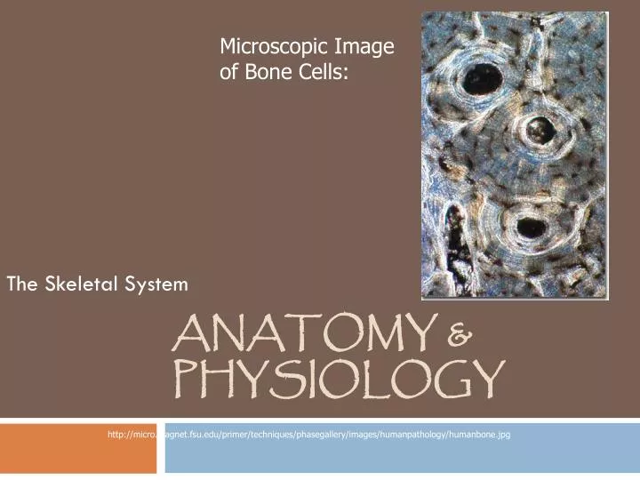
Microscopic Image of Bone Cells:. Anatomy & Physiology. The Skeletal System. http://micro.magnet.fsu.edu/primer/techniques/phasegallery/images/humanpathology/humanbone.jpg. The Skeletal System:. Contains bones , the organs of this system

E N D
Microscopic Image of Bone Cells: Anatomy & Physiology The Skeletal System http://micro.magnet.fsu.edu/primer/techniques/phasegallery/images/humanpathology/humanbone.jpg
The Skeletal System: • Contains bones, the organs of this system • The tissues of this system: bone tissue, cartilage, blood, connective tissue & nervous tissue • Bones, no matter their location or size, have similar functions and structure http://www.nlm.nih.gov/medlineplus/ency/images/ency/fullsize/9065.jpg
Functions of Bones: • Support: provide framework for the body and surround organs • Protection: enclose soft organs • Movement: Along with skeletal muscles and tendons, enable the body to move. • Storage: Within the bone marrow(middle of the bone),Ca++ and P are stored. • Blood Cell Formation: Within the bone marrow, blood cells are formed; a.k.a. hematopoiesis.
Classification of Bones: • There are 206 bones in an adult body. There are 2 types of bone: • Compact Bone: dense • Spongy Bone, a.k.a. Cancellous Bone: open spaces within the bone There are 4 groups of bones: • Long • Short • Flat • Irregular
Types of Bones: http://www.google.com/imgres?
Structure of the Bone: • The diaphysis is the ‘shaft’ of the bone (bone length); mainly compact bone. • The epiphysis are the ends (of long bones); mainly spongy bone. • The periosteum is the bone covering, or membrane. http://www.google.com/imgres?
Structure of the Bone: • Red Marrow forms blood cells; this is found within the epiphysis of some long bones and in spongy bone of flat bones. • Yellow marrow (fat) is found within the medullary cavity. http://www.google.com/imgres?
Microscopic Structure of the Bone: • Osteocytes are mature bone cells. • These are found w/in lacunae(cavities) which form circles calledlamellae. • Lamellae form around Haversian (or osteonic) canals. • Canaliculiallow a ‘transportation’ system for the bone cells (to receive blood and nutrients). • Perforating (or Volkmann’s) canals allow communication to occur.
http://www.web-books.com/eLibrary/Medicine/Physiology/Skeletal/compact_spongy_bone.jpghttp://www.web-books.com/eLibrary/Medicine/Physiology/Skeletal/compact_spongy_bone.jpg
Bone Formation: • Osteomeans bone. • Most bones form from hyaline cartilage. • This process is called ossification. • Bone forming cells are called osteoblasts.
Skeletal Organization: • Look up labeled diagram on p135 in text or online: KNOW THIS FIGURE! • 2 major portions of the skeleton:axial skeleton (bones & cartilage of the head & trunk) and the appendicular skeleton (bones & cartilage of the limbs). • There are 206 bones in the (adult) body.
Axial Skeleton: The Skull: Craniumencloses the brain Includes: • Frontal bone • Parietal Bones (2) • Temporal Bones (2) • Occipital Bone • Sphenoid Bone • Ethmoid Bone http://www.google.com/imgres?
Axial Skeleton: The Skull: Facial Bones, including: • Mandible • Nasal Bones • Maxillary Bones Hyoid Bone: suspended in the midneck above the larynx. http://www.google.com/imgres?
Axial Skeleton: The Vertebral Column (Spine): Contains 33 vertebrae • 9 of these are fused (form 2 bones): • Sacrum and Coccyx(tailbone) • Cervical vertebrae are in the neck region (1st 7) • Thoracic vertebrae are in the trunk (next 12) • Lumbar vertebrae are in the lower back (the last 5) http://www.google.com/imgres?
Axial Skeleton: Thoracic Cage: • Protects the heart, lungs, and major BVs. Includes: 1. Sternum (breastbone) • This is attached to the 1st 7 pairs of ribs. • The heart is posterior to the sternum.
Axial Skeleton: Thoracic Cage: 2. 12 pairs of Ribs: • True ribs are attached to the sternum (1st 7) • False ribs (next 5) • Last 2 pair are a.k.a. ‘floating ribs’ b/c they lack attachment to sternum. • ALL ribs are attached to vertebral column! http://www.google.com/imgres?
Appendicular Skeleton: The Shoulder: Shoulder, or pectoral, girdle contains: • Clavicle (collarbone) • Scapula (shoulder blade) http://www.google.com/imgres?
Appendicular Skeleton: The Upper Limbs: The bones of the upper limb are: • Humerus (arm) • Radius (thumb to forearm) • Ulna (pinky finger to forearm) • Hand: Carpels (wrist), metacarpels (palm), and phalanges (fingers) http://www.google.com/imgres?
Appendicular Skeleton: The Pelvic Girdle: Contains: • Coxal Bones (hip bones) which are composed of the ilium, ischium, and pubis http://www.google.com/imgres?
Appendicular Skeleton: The Lower Limbs: • The thigh bone is a.k.a. the femur. • The leg bones are the tibia (shinbone; larger), fibula (thinner), and patella (kneecap). • The foot contains the tarsal bones (ankle & heel), metatarsals (sole) and phalanges (toes) http://www.google.com/imgres?
Joints: • These are a.k.a.articulations. • This is where 2 or more bones come together. There are 3 types of joints: • Fibrous • Cartilaginous • Synovial
Joints: Fibrous Joints: Cartilaginous Joints: • Between bones that are close together, united by fibrous tissue. • Have limited movement, if any. Sometimes called immovable joints. • Ex. Sutures of the skull • Are shock absorbers & equalize pressure; united by fibrocartilage. • Limited movement. • Ex. Vertebrae
Fibrous Joint: Cartilaginous Joint: http://www.google.com/imgres?
Synovial Joints: Must have: • Articulating cartilage • Articular capsule (there is a membrane) • Joint cavity (synovial fluid) • Ligaments • Many have bursae(flattened sacs of fluid) and tendon sheaths (elongated bursae)
Types of Synovial Joints: • Ball-and-socket jointsallows the most movement: rotational movement, side-to-side, etc. Ex: shoulder or hip. • Condylar jointsallow many motions but not rotational. Ex: between phalanges & metacarpels. • Plane jointsallow sliding & twisting movements. Ex: wrist or ankle.
Plane Joint: Ball & Socket Joint: Condylar Joint: http://www.shockfamily.net/skeleton/GLIDING.JPG http://www.eorthopod.com/images/ContentImages/hip/hip_arthroplasty/hip_arthroplasty_anat01.jpg http://pioneer.netserv.chula.ac.th/~bkritcha/figure/images/condyloid.jpg
Types of Synovial Joints: • Hinge jointsallows planar movement only. Ex: elbow. • Pivot jointsallow rotational movement around a central axis only. Ex: between radius & ulna. • Saddle jointsallow a variety of movements. Ex: between carpal & metacarpal of the thumb.
Hinge Joint: Saddle Joint: http://www.shockfamily.net/skeleton/SADDLE.JPG Pivot Joint: http://www.eorthopod.com/images/ContentImages/elbow/elbow_anatomy/elbow_anatomy02a.jpg http://www.dartmouth.edu/~anatomy/assets/bones/elbow/elbow-supination.jpg
OSSIFICATION TIMETABLE FOR HUMAN OSSIFICATION - LINK http://www.pennmedicine.org/encyclopedia/em_DisplayAnimation.aspx?gcid=000112&ptid=17
Look these up in text or online! Know the following diseases/imbalances: Rickets, fractures, herniated discs, scoliosis, kyphosis & lordosis, bursitis, sprain, arthritis, osteoarthritis, bone spurs, rheumatoid arthritis, ankylosis, and gout, osteoporosis
SPINAL BONE DISORDERS Lordosis Kyphosis
BURSITIS Bursitis is a condition that causes the bursae to become inflamed. This will often need to be treated by protecting the joint from further trauma and keeping the joint rested. Typically bursitis pain will fade after a few weeks if treatment is properly applied, but it is common for those that have suffered from this condition to see flare-ups again in the future. Symptoms of Bursitis Bursitis symptoms may affect the shoulder, elbow, knee, hip or Achilles tendon, which are more common in those over the age of 40. • Pain, particularly when you move the joint or attempt to press on this area • Swelling or redness around the affected area • Stiffness or an ache in the affected area
ARTHRITIS • http://www.pennmedicine.org/encyclopedia/em_DisplayAnimation.aspx?gcid=000092&ptid=17 • Osteoarthritis – most common, associated with aging • Rheumatoid Arthritis • Systemic Lupus Erythematosus • Gout • Childhood Arthritis (Juvenile Idiopathic Arthritis)