Download
1 / 20
200 likes | 309 Views
MLAB 1415: Hematology Keri Brophy-Martinez. Chapter 26: Lymphoid Malignancies Part Two. Lymphomas.

E N D
MLAB 1415: HematologyKeri Brophy-Martinez Chapter 26: Lymphoid Malignancies Part Two
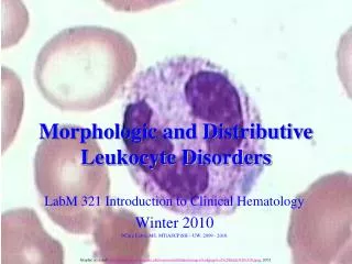
Lymphomas • These are a heterogeneous group of diseases that arise from an overproliferation of abnormal cells of the lympoid tissue (lymphocytes, histiocytes, and reticulum cells). The spilling of these cells into the peripheral blood results in a leukemic phase of the disease.
Hodgkin’s Lymphoma • Etiology and clinical features • Probable cause is Epstein-Barr virus. • Diagnosed between 15 and 35 years of age; also found in over 50 population • Nonpainful lymph node swelling
Hodgkin’s lymphoma • Pathology • Characteristic cell is the Reed-Sternberg • Giant size (up to 45µm in diameter) • Abundant acidophilic cytoplasm • Multinucleated or polylobated nucleus • Gigantic nucleoli
Hodgkin’s lymphoma • Staging of Hodgkin’s • Stage I - single lymph node region or single organ • Stage II - two or more lymph node regions on the same side of the diaphragm • Stage III - involvement of lymph nodes on both sides of the diaphragm • Stage IV - disseminated into other tissues and organs including bone marrow
Hodgkin’s lymphoma • Treatment and prognosis • Radiation of localized involvement • Chemotherapy • Combination of above • With early diagnosis, long-term disease-free survival is seen in about 75% of cases.
Non-Hodgkin’s Lymphoma • Cause is unknown at this time. Predisposing factors seem to be chemicals, ionizing radiation and certain viruses. Reed-Sternberg cells are NOT present. • The types of non- Hodgkin’s lymphoma reflect the developmental stages of lymphocytes. • Each type of lymphoma can be viewed as a lymphocyte arrested at a certain stage of development and transformed into a malignant cell. • 85% B cell origin, the rest T or null cell.
Burkitt Lymphoma • Endemic to Africa • 1/3 of all non-African pediatric lymphomas • Found in immunocompromised patients, particularly AIDS patients • Tumor growth rate is the highest of any tumor with growth doubling each day. • Rapid growth and tumor cell death results in “starry sky” appearance of the biopsy caused by macrophages cleaning up the dead cells. • Characteristic overgrowth of facial bones in the African variety and abdominal mass in the non-endemic variety. • Cytogenetic translocation t(8:14)
Burkitt Lymphoma • Overgrowth of facial bones
Plasma Cell Disorders • Disorders that do not involve lymph nodes • Secrete monoclonal immunoglobulin into the serum and /or urine • Disorders • Multiple Myeloma • Plasmacytoma • Primary amloidosis • Heavy Chain disease • Monoclonal gammopathy of undetermined significance
Multiple myeloma • This is a disorder in which there is overproduction of abnormal plasma cells which are the final stage in the development of B lymphocytes.
Etiology • 50% greater risk for men than women • Risk increases with age; rare under 40 • Median age 65 years old • Suspected cause is chronic stimulation of the immune system from environmental sources. • Ionizing radiation • Viruses
Features • Expanding plasma cell mass in bone marrow causes pancytopenia and destruction of the bone cortex. This is painful because nerves get stretched. • Formation of tumors causes lytic bone lesions Bone lesions
Features • Increased production of immunoglobulin heavy and light chains (monoclonal gammopathy) • Heavy chains: IgG, IgA, IgD, IgE, IgM • Light chains: kappa, lambda • Most common type of multiple myeloma is increased production of IgG. • Hyperviscosity syndrome • Excess immunoglobulin causes viscous blood which sludges and causes fluid congestion. • Bence-Jones protein • Light chains spill into the urine and can be detected by lab test. • Causes kidney damage
Lab Findings • CBC and peripheral smear • Red cells form characteristic rouleaux formation (resemble stacked coins) • Plasma cells may be seen in advanced cases • The presence of significant numbers of plasma cells on peripheral blood smear constitutes plasma cell leukemia • ESR • Increased - serum protein causes red cells to stick together and fall faster • Bone marrow • Increased number of plasma cells which form “sheets” • Chemistry studies • Increased BUN and creatinine (kidney tests) • Increased calcium • Increased LDH
Multiple myeloma • Treatment • Chemotherapy • Radiation for localized areas • Bone marrow transplant for younger patients
Waldenström’s macroglobulinemia • Overproduction of monoclonal IgM by plasma cells • Clinical features • Usually presents when patients are in their 70's. • Hyperviscosity syndrome is common. • Blurred vision • IgM interferes with platelet function and bleeding problems occur. Characteristic bruising is calledcryoglobulinemic purpura • Cryoglobulins precipitate on exposure to cold
References • http://www.itriagehealth.com/wl/disease/burkitt-lymphoma-%28lymph-node-tumor%29#wrapperTop • McKenzie, S. B., & Williams, J. L. (2010). Clinical Laboratory Hematology . Upper Saddle River: Pearson Education, Inc.