Download
1 / 28
300 likes | 374 Views
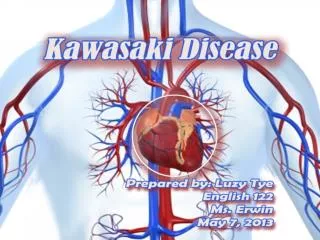
Learn about Kawasaki disease, a systemic vasculitis affecting young children, its clinical manifestations, complications, and imaging studies for diagnosis and management.

E N D
Presentation – Patient 1 • 10 years old male with Kawasaki Disease • Possible mildly ectatic posterior descending coronary artery on Echo. • CTA was performed. • Heart rate control • Contrast enhanced axial images of the coronary arteries • Multiplanar and 3D reformats
Presentation – Patient 2 • 13 years old male with Kawasaki Disease and known coronary aneurysm presents for CTA. • Heart rate control • Contrast enhanced axial images of the coronary arteries • Multiplanar and 3D reformats
Presentation – Patient 3 • 13 years old female with Kawasaki Disease and known coronary aneurysm diagnosed on Echocardiogram presents for CTA.
Kawasaki Syndrome • AKA – Mucocutaneous Lymph Node Syndrome • First characterized in 1967 by Dr. Tomisaku Kawasaki of Tokyo • Multisystem vasculitis • Self limited • Leading cause of acquired heart disease in the US.
Epidemiology • Syndrome of young children • >80% are < 4y/o • Most are between 1-2y/o • Very rare > 8y/o • Males > females • Japanese and Korean ancestry > other ethnicities • 4,000-8,000 cases/yr
Etiology • Unknown • Leading hypothesis • Infectious with immune-mediated reaction • No single etiologic agent identified
Clinical Manifestationand Diagnosis • No recognized prodrome • Acute onset • Begins with fever • Other symptoms begin within 2-5 days • Syndrome • Fever for 5d + 4/5 symptoms • Fever for 5d + 3/5 symptoms + coronary artery abnormalities
Signs/symptoms • Nonpurulent bilateral conjunctivits • Oropharyngeal changes • Erythema, redness, cracking, peeling, injected pharynx, strawberry tongue • Polymorphous erythematous rash • Most evident with fever • Primarily on trunk, may be pruritic • Cervical lymphadenopathy • Often unilateral • One node >1.5cm • Feet/Hand changes • Erythema (palms/soles), edema, induration, desquamation
Signs/Symptoms • Cardiac findings • Leading cause of morbidity and mortality • May involve pericardium, myocardium, endocardium, valves, coronary arteries
Signs/Symptoms • 20-25% of untreated pts develop coronary artery abnormalities/aneurysms • Within 4 weeks of onset • Dilation can be detected at 10d • Peak 18-25d • Giant aneurysm >8mm • Greatest risk thrombosis, stenosis, MI
Complications • Cardiac manifestations can be deadly • Most deaths occur between 2-12 weeks post illness onset • Coronary aneurysm thrombosis – MI • Aneurysm rupture • Myocarditis - CHF
PredictingCoronary Artery abnormalities • Harada score • White blood cell count > 12K • Platlet count < 350K • CRP >3+ • Hct < 35% • Albumin < 3.5 g/dL • Age < 12 months • Male sex • Used to determine management
Treatment • Decrease inflammation • Prevent thrombosis • Overall goal • Protect the myocardium and coronary arteries • Modes of treatment • IVIG • Steroids • TNF –alpha antagonists • Aspirin • abciximab
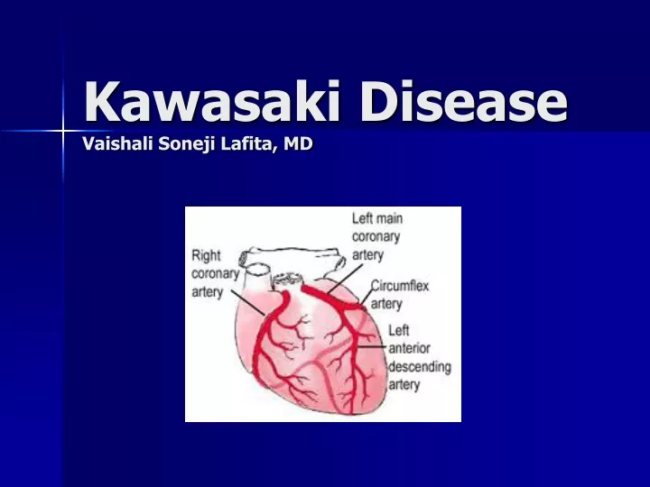
Cardiac findings : Imaging • Echocardiography • Unclear sensitivity and specificity • Coronary Angiography • Intravascular Ultrasound (IVUS) • Transesophageal echocardiography • MRA • CTA
Cardiac findings :Imaging • Echocardiogram • 1st at time of diagnosis • Number, location and classification of aneurysms • Small, medium or giant • Repeat echo 2-3 and 6-8 weeks after onset of the disease • If both normal – no further echos needed • If abnormal • Severity determines frequency and type of follow-up
Management of Aneurysms • Small to medium:long term Aspirin, no activity restriction, stress test in older children if suggests stenosis→ coronary angiography • Multiple, small to medium or giant: Aspirin with/without warfarin, >10y stress test with myocardial perfusion scan if coronary obstruction→ bypass grafting, angioplasty
Summary • Self limited systemic vasculitis • Leading cause of acquired heart disease in the US. • Morbidity and mortality from cardiac involvement. • Imaging plays critical role in diagnosis and management of cardiac involvement.
References • J. W. Newburger, M. Takahashi, M. A. Gerber, M. H. Gewitz, L. Y. Tani, J. C. Burns, S. T. Shulman, A. F. Bolger, P. Ferrieri, R. S. Baltimore, W. R. Wilson, L. M. Baddour, M. E. Levison, T. J. Pallasch, D. A. Falace, and K. A. TaubertDiagnosis, Treatment, and Long-Term Management of Kawasaki Disease: A Statement for Health Professionals From the Committee on Rheumatic Fever, Endocarditis, and Kawasaki Disease, Council on Cardiovascular Disease in the Young, American Heart AssociationPediatrics, December 1, 2004; 114(6): 1708 - 1733. • http://www.emedicine.com/EMERG/topic811.htm • http://www.cdc.gov/ncidod/diseases/kawasaki/index.htm • http://www.americanheart.org/presenter.jhtml?identifier=11163