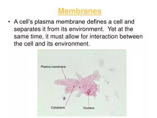
Download
1 / 20
200 likes | 365 Views
Membranes. Spheres of Hydration Around Two Charged Ions. Salt ( NaCl ) dissolves in water and separates into Na + and Cl -. Fatty Acids . Carbon backbone Carboxyl group ( - COOH) Unsaturated One or more double bonds in backbone Saturated

E N D
Spheres of Hydration Around Two Charged Ions Salt (NaCl) dissolves in water and separates into Na+ and Cl-
Fatty Acids • Carbon backbone • Carboxyl group (- COOH) • Unsaturated • One or more double bonds in backbone • Saturated • All single bonds in backbone
Triglycerides • Neutral fats • Three fatty acids and a glycerol • Condensation reaction • Body’s mostabundant lipid • Functions: • Energy reservoir • Insulation
A Phospholipid: Lipid tails Phosphate head
sterol backbone In-text, p. 41 Sterols • Sterols • No fatty acid tails • Four carbon rings • Promote fluidity in eukaryotic cell membranes • Example: cholesterol
Plant vs. Animal Cells: • Animal cells have: • Centrioles • Gap, adhering and tight junctions • Plant cells have: • Chloroplasts • Cell walls • Plasmodesmata
Plant Cell Wall: • Cell secretions form lamella • Plasmodesmata (channels) • Primary and Secondary walls
Cell-to Cell Junctions: • Plants • Plasmodesmata • Animals • Tight Junctions • Adhering Junctions • Gap Junctions
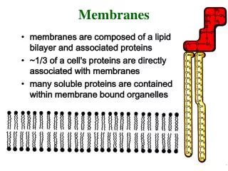
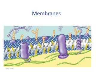
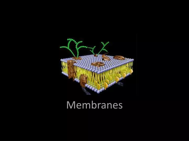
Cell Membrane Structure: • Also called a • plasma membrane • phospholipidbilayer • Fluid mosaic • Major components: • Phospholipids • Proteins (4 types) • Sterols http://www.funhousefilms.com/cellmemb.jpg
hydrophilic head one layer of lipids hydrophobic tails one layer of lipids Phospholipid Bilayer: Fig. 4.3, p. 54
LIPID BILAYER cytoplasm extracellular fluid
Surface Markers and Sterols: http://www2.bioch.ox.ac.uk/~drick/images/mann.jpg
The Cytomembrane System: • Organelles in which lipids are assembled and proteins are produced and modified and then secreted. The following organelles are involved: • Endoplasmic reticulum • Ribosomes • Golgi bodies • Vesicles
assorted vesicles Some vesicles form at the plasma membrane, then move into the cytoplasm. These endocytic vesicles might fuse with the membrane of other organelles or remain intact, as storage vesicles. Other vesicles bud from ER and Golgi membranes, then fuse with the plasma membrane. The contents of these exocytic vesicles are thereby released from the cell. 5Vesicles budding from the Golgi membrane transport finished products to the plasma membrane. The products are released by exocytosis. Golgi body 4Proteins and lipids take on final form in the space inside the Golgi body. Different modifications allow them to be sorted out and shipped to their proper destinations. smooth ER 3 Vesicles bud from the ER membrane and then transport unfinished proteins and lipids to a Golgi body. 2 In the membrane of smooth ER, lipids are assembled from building blocks delivered earlier. rough ER 1 Some polypeptide chains enter the space inside rough ER. Modifications begin that will shape them into the final protein form. DNA instructions for building polypeptide chains leave the nucleus and enter the cytoplasm. The chains (green) are assembled on ribosomes in the cytoplasm. EndomembraneProtein Synthesis Fig. 4.13, p. 64
Transport Proteins: • Allow water soluble molecules through themselves • They are transmembrane • Open channels, gated channels, carriers, and pumps Carrier Protein Channel Protein Symport Antiport
Receptor Proteins: • Bind hormones
Recognition Proteins: • Identify cells • Help guide cells into tissues • Cell to cell recognition and coordination
cytoskeletal protein (e.g., clathrin) ADHESION PROTEIN open channel gated channels (open and closed) active transport RECEPTOR PROTEIN RECOGNITION PROTEIN LIPTD BILAYER TRANSPORT PROTEINS Adhesion Proteins: • Help cells stay connected Fig. 5.21, p. 92