Download
1 / 16
380 likes | 1.11k Views
Electrical Activity of the Heart. Chloe, Chanel, Raj. Conducting System. AV node Sa node Purkinje fibers Bundle of his. SA node. S inoatrial node Specialized group of cardiac muscle cells that don’t contract located in the right atrium of the heart Adapted to automatically

E N D
Electrical Activity of the Heart Chloe, Chanel, Raj
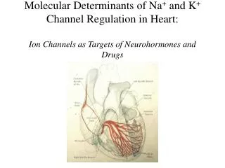
Conducting System • AV node • Sa node • Purkinje fibers • Bundle of his
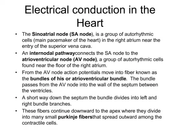
SA node • Sinoatrial node • Specialized group of cardiac muscle cells that don’t contract • located in the right atrium of the heart • Adapted to automatically generate impulses • generator of heart rhythm • Basically the pace maker
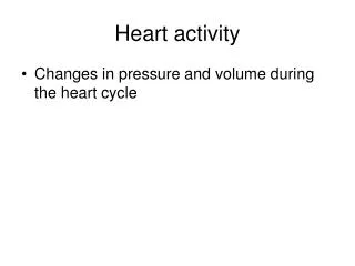
Function • It triggers the electrical impulses that are carried around to other heart cells • The electrical impulses cause the atria to contract at a rate of 60 to 100 beats per minute • Signals atria to contract, passes the blood to bottom • Causes contraction
AV node • Atrioventricularnode • electrically connects atrial and ventricular chambers • specialized tissue between the atria and the ventricles of the heart • Also automatically generates impulses- cause contraction
Functions • begins when it gets an electrical impulse from the SA node • The rhythm of the heart contractions is set by the AV node. It delays the impulse by about 0.12 seconds. • This is delay is very important as it helps the atria eject the blood in them into the ventricles before the contraction of the ventricles. • The delay also helps in protecting the ventricles from atrial arrhythmia (type of arrhythmia). • Sends the signal it receives to the fibers below, opens the semilunar, blood is forced out • http://www.youtube.com/watch?v=0xUifyll2Oc • http://www.youtube.com/watch?v=lIQXzgesdDg
Purkinje fibers • located in the inner ventricular walls of the heart, just beneath the endocardium. • consist of specialized cardiomyocytes that are able to conduct cardiac action. • allow the heart's conduction system to create synchronized contractions of its ventricles, and are therefore essential for maintaining a consistent heart rhythm.
Bundle of his • collection of heart muscle cells, known as cardiomyocytes, specialized for electrical conduction that transmits electrical impulses. • The fascicular branches then lead to the Purkinje fibers which provide electrical conduction to the ventricles, causing the cardiac muscle of the ventricles to contract at a paced interval. • Basically directs orders to branches that allow a Heart to contract. These branches that the His bundle directs, are the Purkinje fibers.
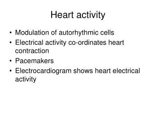
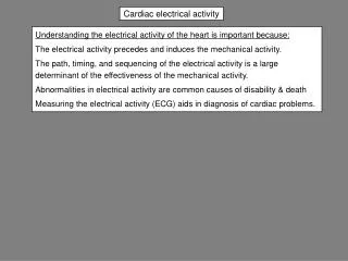
EKG- What is it? • Electrocardiogram • Checks for problems with the electrical activity of your heart • Natural electrical system causes the heart muscle to contract and pump blood • Translates electric activity in heart into line tracings on paper • Spikes and dips in line tracings called waves
Why Is it done? • Find cause of unexplained chest pain • Find cause of symptoms of heart disease • Check thickness of heart chambers (hypertrophied) • Check effectiveness of medicines • Check overall health of heart
How it’s done • EKG paste placed between the electrodes and your skin to help conduction of electrical impulses • Several electrodes are attached to the skin and hooked to machine • Machine traces heart activity on paper
Works Cited • http://www.beltina.org/health-dictionary/atrioventricular-av-node.html • http://biology.about.com/library/organs/heart/blatrionode.htm • http://www.wisegeek.com/what-are-purkinje-fibers.htm • http://medical-dictionary.thefreedictionary.com/Purkinje+fiber • http://www.webmd.com/heart-disease/electrocardiogram • http://www.nhlbi.nih.gov/health/health-topics/topics/ekg/ • http://www.beltina.org/pics/atrioventricular__av__node.gif