Download
1 / 30
300 likes | 318 Views
Explore the intricate relationship between Curcumin and Diphtheria Toxin receptor for enhanced drug delivery in cancer therapeutics. Investigate binding interactions and cellular uptake mechanisms with fluorescence spectroscopy and MTT assays.

E N D
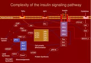
Complexity of Signaling Networks Old Protein, New Tricks Biplab Bose
Diphtheria Toxin Exotoxin of Corynebacterium diphtheriae.
HB-EGF: DT Receptor Heparin-binding epidermal growth factor (EGF)-like growth factor Soluble & Membrane bound • In Oncogenesis: • Increases proliferation • Induction of migration and invasion • Promotion of angiogenesis • Normal Function • Cell proliferation • Developmental process • Wound-healing Overexpressed in tumors: Pancreatic, Liver , Gastric and Glioma
Receptor Binding Domain of DT (RDT) R-domain of Diphtheria Toxin Diphtheria Toxin PDB ID: 1F0L
Cloning of RDT SOURCE: Full Length DT cloned in pET-22b
Expression & Purification of RDT • E. coli BL21(DE3) • Induction at 28 0C ,1 mM IPTG. • Purification by His-Trap Column. Western Blot using anti-His Ab SDS-PAGE of purified RDT
RDT binds to HB-EGF Solid Phase ELISA: • HB-EGF Coated on 96-well ELISA plate. • Detection: anti-His Ab followed by anti-mouse Ab
RDT Binds to Cell Surface HB-EGF Immunofluorescence: • Cell line: U-87 MG. • Detection: anti-His Ab followed by FITC-Conjugated anti-mouse Ab
Binding Affinity of RDT SPR, Biacore X-100 • Single cycle Kinetics. • CM5 Chip, HB-EGF immobilized
Can a Drug Bind to RDT ? • blue = hydrophilic and • orange red = hydrophobic.
Water quenches curcumin fluorescence. Curcumin: a Good Probe Fluorescence of Curcumin depends upon environment
Curcumin: a Good Probe Curcumin binds to some proteins Protein binding increases fluorescence of Curcumin
Docking of Curcumin on RDT Docking server: SwissDock Docking Criteria: Blind, no flexibility for RDT. RDT Structure of Curcumin (from ZINC) Source: R-domain of B-chain of 1FOL
Docking of Curcumin on RDT A potential binding pose Surface Diagram Ribbon Diagram • Blue = Hydrophilic • Orange red = Hydrophobic.
Docking of Curcumin on RDT Important interactions between RDT and curcumin:
Curcumin Binds to RDT Fluorescence spectroscopy: • Curcumin (10 µM) in PBS, Molar ratio of Curcumin:Protein (10:1) • Incubation at 4 0C, 2 hr • Excitation at 430 nm.
Curcumin Binds to RDT Fluorescence spectroscopy: • Curcumin (10 µM) in PBS, with RDT varied (0 to 2 µM) • Incubation at 4 0C, 2 hr • Excitation at 430 nm.
Curcumin Binds to RDT Time-resolved fluorescence spectroscopy: • Excitation at 405 nm • Curcumin (10 µM) in PBS, Molar ratio of Curcumin:Protein(10:1) • Decay measured in ns/channel. • Exponential component analysis.
Curcumin-RDT enhances accumulation of curcumin Cell Line: U-87 MG Curcumin (2 µM); molar ratio curcumin:protein (10:1) 20X
Curcumin-RDT increases cellular uptake of curcumin HPLC: • Cell Line : U-87 MG • Curcumin:1 µM; • Curcumin-protein • (molar ratio:10:1) • Incubation at • 37 0C, 2 hr, Serum. • C18 column; • methanolic cell extract ** * * * No significant difference between these two (p = 0.143); **significantly different from others (p < 0.001). One-way ANOVA with pairwise comparison
Curcumin-RDT potentiate curcumin MTT assay: • Cell line: U-87 MG • RDT: 0.1 µM • Incubation at 37 0C, • 72 hr, Serum-Free Significant difference between Curcumin and Curcumin-RDT: 2-way ANOVA, p<0.001
Effect of Curcumin-RDT is not synergistic MTT Assay • Cell line: U-87 MG • RDT: 0.1µM; Curcumin:20 µM; • Curcumin-RDT: 20 µM:0.1 µM; ** Significantly different from other treatment groups (p < 0.001)
Effect of Curcumin-RDT on cell cycle Flowcytometry • Cell line: U-87 MG • RDT: 0.1 µM; Curcumin:20 µM; • Curcumin-RDT: 20 µM:0.1 µM; • 48 hr.
Curcumin-RDT enhances apoptosis Flowcytometry • Cell line: U-87 MG • RDT: 0.1 µM; Curcumin:20 µM; • Curcumin-RDT: 20 µM:0.1 µM; • Incubation at 37 0C, 48 hr. PI Annexin V