Download
1 / 54
540 likes | 778 Views
MAXIMIZING THE UTILITY OF THE PRIME ECG. James Hoekstra, MD Professor and Chairman Department of Emergency Medicine Wake Forest University. SO HOW DO YOU READ THIS DAMN THING ANYWAY??. Patient Selection Evaluating the PRIME ECG results. PRIME ECG Patient Selection.

E N D
MAXIMIZING THE UTILITY OF THE PRIME ECG James Hoekstra, MD Professor and Chairman Department of Emergency Medicine Wake Forest University
SO HOW DO YOU READ THIS DAMN THING ANYWAY?? • Patient Selection • Evaluating the PRIME ECG results
PRIME ECG Patient Selection • The PRIME ECG does not replace the screening ECG • Too time intensive • Too expensive • PRIME sensitivity and specificity was determined from high risk subsets of patients • Low risk chest pain patients will result in high false positive rates, just like the 12 lead ECG
PRIME ECG Patient Selection • High Risk Patients • High index of suspicion for evolving STEMI: Serial PRIME • ST Depression • Abnormal but nonspecific ECG, BBB, LVH • Troponin Positive (after TnI or TnT comes back) • TIMI 2+
PRIME ECG Patient Selection • Should not be used to screen for “safe to go home” screening scenarios • Should not be used in “observation unit” patients (TIMI 0-1) • Should be used in “admissions,” in serial fashion, especially if high risk
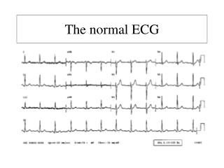
Reading the PRIME ECG • Regimented Reading • Assure a quality recording • Stepwise approach to reading • Match the ECG to the Patient • Like the 12 Lead ECG, when in doubt, go back to the clinical scenario
Reading the PRIME ECG • Regimented Reading Approach • Low Quality Screen • Assisted Beat Markings • 80 Lead Screen • 4 MAP View • STO Filter View • Computerized Reading
PRIME ECG Case Study:54 yo Male with Chest Pain • Arrives in the ED with chest pain that had been constant since 11 AM. • Pain is described as intense, midline substernal, radiating to the left arm, associated with shortness of breath and nausea. It began with light walking. It feels like his prior MI pain • Pain 8/10 on arrival, in mild distress
54 year old Male with CP (PMH) • PMH: CAD, MI • Prior stent placed for MI • SH: Smoker • FH: Noncontributory • Meds: ASA. Noncompliant with other medications
54 year old Male with Chest Pain (ECG) • Normal Sinus Rhythm at 72 bpm • ST depression anteriorly • Tall R waves anteriorly
54 year old Male with CP (Ancillary) • Chest Xray: Normal • Initial Cardiac Markers: • CK 37, MB ,1.0 • TnI < 0.05 • Renal Function Normal • Hb 14 • TIMI 5
54 year old Male with CP (PRIME) Low Qual • STEP 1: LOW QUALITY REVIEW
54 year old Male with CP (PRIME) Rate, Axis, Intervals • STEP 1: LOW QUALITY REVIEW 80 Lead Toggle Low Quality Lead
54 year old Male with CP (PRIME) Hit Analyze Button • STEP 2: ACCEPT BEAT MARKINGS If OK, Accept
54 year old Male with CP (PRIME) STO Takeoff End of T • STEP 2: ACCEPT BEAT MARKINGS Start of QRS
54 year old Male with CP (PRIME) Step 3: Scroll Through the 80 Leads 80 Lead View Button
54 year old Male with CP (PRIME) 4 View Torso View • Step 4: MAP 4 View (Torso) Isolate PQRST Max ST Deviation
54 year old Male with CP (PRIME) STO • Step 5: ST0 Filter
54 year old Male with CP (PRIME) Rotate • Step 5: Rotate Filter
54 year old Male with CP (PRIME) • Step 6: Read the Analysis: Post MI
54 year old Male with CP (ED Course) • Cardiology consulted for possible acute posterior MI • Cardiologist saw pt in the ER • Pain reduced, workup complete, decision is made to admit to CCU for medical management pre-cath (treat as NSTE ACS)
54 year old Male with CP (CCU Course) • Second set of markers elevated with CKMB 37, TnI 6.6 • Pain continues, 2-3/10, despite maximal medical management • Patient taken to cath lab for urgent PCI
54 year old Male with CP (Cath Results) • 99% thrombotic lesion of the proximal first obtuse marginal off the circumflex • Remainder of circumflex without stenoses • LAD and RCA with mild lumenal irregularities, none more than 20% obstructive • LVEF 50%
54 year old Male with CP (Course) • Patient underwent PCI with taxus stent of the proximal obtuse marginal 99% lesion with good result. • Discharged home on hospital day 5 • Final diagnosis: Acute Posterior MI
PRIME ECG Case Study:53 yo Male with Chest Pain • Arrives in the ED with chest pain that had been stuttering for the past 12 hours • Pain is described as dull, substernal, associated with shortness of breath and nausea • Pain 2/10 on arrival, in no distress
53 year old Male with CP (PMH) • PMH: HTN, CVA, Seizures, CAD, Ashma • EF 40% secondary to past MIs (anatomy unclear due to cath at outside hospital) • SH: Smoker 1 ppd X 30 years • FH: Noncontributory • Meds: ASA, Lipitor, Lisinopril, Lamictal, Albuterol, Lopressor, Neurontin
53 year old Male with Chest Pain (ECG) • Normal Sinus Rhythm at 79 bpm • Diffuse nonspecific ST/T wave changes anteriorly • Early R wave progression anteriorly
53 year old Male with CP (Ancillary) • Chest Xray: Mild cardiomegaly, no CHF • Initial Cardiac Markers: • CK 221, MB 5.3 • TnI < 0.05 • Renal Function Normal • Hb 14
53 year old Male with CP (ED Course) • Cardiology consulted for acute anterior MI • Cath Lab Activated • Cardiologist at Bedside, agrees with PRIME interpretation • Angiogram performed
53 year old Male with CP (Cath Results) • LAD with 95% mid lesion • Critical stenosis of first diagonal branch • 85% circumflex 2nd OM lesion • 70% circumflex 3rd OM lesion • 75% lesion of PDA off the RCA • Apical hypokinesis and LVEF 40%
53 year old Male with CP (Course) • Cardiothoracic Surgery consulted for CABG due to triple vessel disease • Felt not to be surgical candidate due to prior CVA • Day 2 underwent stenting of LAD 95% lesion, without complication • Peak Troponin 0.10 • Discharged home on hospital day 5 • Diagnosis: Unstable Angina
PRIME ECG Case Study:62 yo Male with Chest Pain • Arrives in the ED by EMS with chest pain that has been intermittent for 2 days, and constant for the last 2 hours. • Pain is described as substernal, radiating to the left arm, associated with shortness of breath and nausea. He has been taking NTG without relief • Pain 8/10 on arrival, in moderate distress
62 year old Male with CP (PMH) • PMH: CAD, MI, DM, HTN, CHF, Neuropathy • PSH: CABG, Pacemaker • SH: Nonsmoker • FH: MI • Meds: ASA, Clopidogrel, Glucophage, Lantis, Lopressor, Lisinipril, Lasix, Lipitor, Neurontin
62 year old Male with Chest Pain (ECG) • Normal Sinus Rhythm at 72 bpm • Bifascicular Block RBBB and LAHB • Diffuse ST depression over anterior leads
PRIME Obtained 290
62 year old Male with CP (ED Course) • Cardiology consulted for possible acute posterior MI • Cardiologist at bedside. Agrees with PRIME Reading • Patient offered PCI, but initially not willing to undergo cath. Agreed later after second set of TnI elevated at 6.1
62 year old Male with CP (Cath Results) • LAD, RCA, and circumflex all occluded from the native circulation. • LIMA graft to LAD patent, distal LAD 80% stenosis • Saphenous graft to the RCA patent without stenoses • Saphenous graft to the circumflex occluded at the distal anastomosis • LVEF 30%
62 year old Male with CP (Course) • Patient underwent PCI with taxus stent of the distal saphenous graft to the circumflex, with good results. • Patient admitted to the CCU post procedure • Peak CKMB >80, Peak TnI >30 • Discharged home on hospital day 5 • Final diagnosis: Acute Posterior MI
OCCULT – MI TRIAL • Mitchell Krucoff, MD and James Hoekstra, MD - Co-chair • Heartscape - Sponsor • Steering Committee • DCRI - Prime ECG Core Laboratory • PERFUSE Angiographic Core Lab • Cardiovascular Clinical Studies -CRO
OCCULT – MI Trial Sites Bay State Medical Center Occult-MI study 1400 patients 10 + sites Columbia-Presbyterian William Beaumont Thomas Jefferson Cleveland Clinic Wake-Forest UC-Davis University of Cincinnati Duke Medical University of South Carolina * Tallahassee Heart & Vascular Institute * Tampa General
Inclusion Criteria • Able to consent • >39 years old • Has access to a working telephone and the ability to hear by phone • Non-trauma associated ACS symptoms beginning 12 hours or less before presentation • Chest pain and at least one of the following: (i) ECG abnormality; (ii) known CAD; (iii) at least 3 coronary risk factors for CAD (including: family history, current or treated hypertension, hypercholesterolemia or treatment for it, diabetes mellitus and/or subject is a current smoker).
Exclusion Criteria • Symptoms > 12 hours • Prior 12-lead STEMI within the past 48 hours • Hemodynamic instability • Cardiogenic shock • Pulmonary edema (Killips class 3: overt failure with 1/3 of lung fields) • Recent trauma.
Methods: Data Capture • Prospective, cohort study of PRIME ECG • Participants blinded to PRIME result • Brief medical history (vital signs, height, weight, cardiopulmonary exam, concomitant medications) • 12-L and PRIME SERIAL recordings – near simultaneous at enrollment, simultaneous at change of symptoms or at least one additional recording within 3 hours; with pain assessment at time of each recording • Clinical labs including cardiac markers – must include troponin [ I or T ] • TIMI risk assessment • Interventional therapies • Angiographic films • Results of: stress MPI, echocardiography, and SPECT scan during index ED visit / hospitalization • 30 day f/u for MACE
Primary Endpoint DTST for Prime-only STEMI subjects vs. STEMI subjects. DTST will be measured in minutes, from the time stamped on the ED intake sheet to the time of sheath insertion in the cardiac catheterization laboratory.
Secondary Endpoints • 10+ endpoints of clinical and economic factors • Sub-group analyses between Prime-only STEMI, STEMI, non-STEMI • Clinical factors analyzed will include: 30-day MACE rates, AMI detection, ACS detection, angiographic determination of arterial stenosis/occlusion, revascularization rates, medical therapy regimens
PRIME ECG Case: OCCULT MI • 6/10 Pain • ECG with ST depression only in anterior and lateral leads • Posterior leads OK • PRIME ECG applied 001-046
PRIME ECG Case Resolution • Door to cath time 486 minutes • TnI 186X ULN • Final Dx NSTEMI
PRIME ECG Case: OCCULT MI ECG with nonspecific findings TIMI 2 Pain 2/10 on arrival, PRIME Applied 003-0148