Download
1 / 26
270 likes | 460 Views
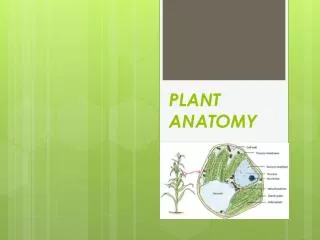
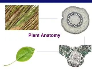
Plant Anatomy Pictures. Plant tissues fit into 3 main categories: Dermal tissues : epidermis, trichomes (“hairs”), guard cells, root hairs Ground tissues : parenchyma (with chloroplasts = chlorenchyma), sclerenchyma, collenchyma

E N D
Plant tissues fit into 3 main categories: Dermal tissues: epidermis, trichomes (“hairs”), guard cells, root hairs Ground tissues: parenchyma (with chloroplasts = chlorenchyma), sclerenchyma, collenchyma Vascular tissues: xylem (dead at maturity: tracheids and vessel elements), phloem (living: sieve tube cells [no nuclei], companion cells [with nuclei])
Stem tip: apical meristem divides, adding on new cells; cells mature into various tissues.
Region of cell division (mitosis) in a root tip. Cells with chromo-somes can be seen in stages of mitosis.
Cross-section: young dicot stem with ring of vascular bundles
Cross-section: young dicot stem with ring of vascular bundles
Cross-section: woody stem showing 3 years of secondary growth. Note pith at center. Dark (reddish) ring is the bark containing a layer of living phloem and outer dead cork.
Monocot stem section showing scattered vascular bundles and enlargement of single vascular bundle (“monkey face”).
Cross-section of a root showing branch root formed by pericycle.
Epidermal cells of root showing formation of root hairs from single cells.